Kyung Min Nam1, Ayhan Gursan1, Alex Bhogal1, Jannie Wijnen1, Dennis Klomp1, Jeanine Prompers1, and Arjan D. Hendriks1
1University of Medical Center Utrecht, Utrecht, Netherlands
1University of Medical Center Utrecht, Utrecht, Netherlands
Deuterium Echo-Planar Spectroscopic Imaging (DEPSI)
is proposed as a way to increase the spatial and temporal resolution of 7T
deuterium metabolic imaging (DMI). The sensitivity was high enough to monitor
in-vivo liver glucose metabolism with 20 mm3 resolution and a scan
time of < 10min.
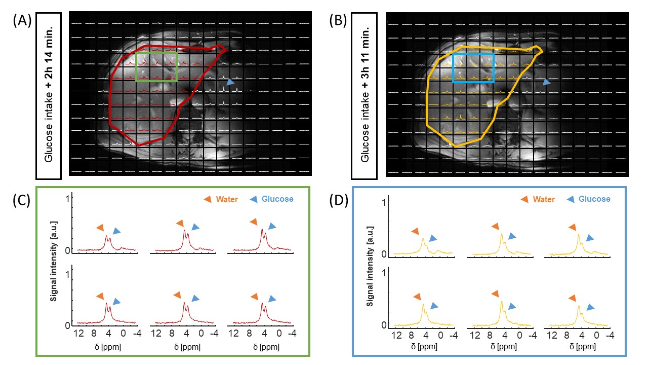
Fig. 4: 3D
DEPSI scans of the liver, acquired respectively two hours (A and C) and 3 hours
(B and D) after intake of deuterated glucose (glucose). In the MRI-DMI overlays
(A and B), the liver contours are highlighted. Selected voxels in the liver (C
and D) show both the glucose peak (blue marker) and the water peak (orange
marker). It can be seen that the glucose peak decreases over time. Note that
the glucose signal in the stomach (A, blue marker) can also be detected and
that it is much higher 2 hours after intake than 3 hours after intake (B).
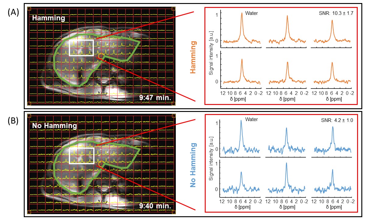
Fig. 3: 3D DEPSI datasets of the human liver at natural
abundance, acquired with Hamming weighting (A) and without Hamming weighting
(B). The left panels show overlays of the 1H MRI images (Dixon) and
the 2H spectra. The right panel highlights 6 voxels located in the
liver. The average SNR of the deuterated water signal in the ROI (white box) is
2.4 times higher for the Hamming weighted DEPSI acquisition.