Yaewon Kim1, Brian T. Chung1, Jeremy W. Gordon1, Adam W. Autry1, Chou T. Tan2, Chris Suszczynski2, Susan M. Chang3, Yan Li1, Duan Xu1, and Daniel B. Vigneron1,3
1Department of Radiology and Biomedical Imaging, University of California, San Francisco, CA, United States, 2ISOTEC Stable Isotope Division, MilliporeSigma, Merck KGaA, Miamisburg, OH, United States, 3Department of Neurological Surgery, University of California, San Francisco, CA, United States
1Department of Radiology and Biomedical Imaging, University of California, San Francisco, CA, United States, 2ISOTEC Stable Isotope Division, MilliporeSigma, Merck KGaA, Miamisburg, OH, United States, 3Department of Neurological Surgery, University of California, San Francisco, CA, United States
A new method for hyperpolarized [2-13C]pyruvate MRI with volumetric and dynamic EPI of the human brain with metabolite-specific in-plane resolution was developed and applied in healthy volunteers to investigate both TCA and glycolytic metabolism in the same acquisition.
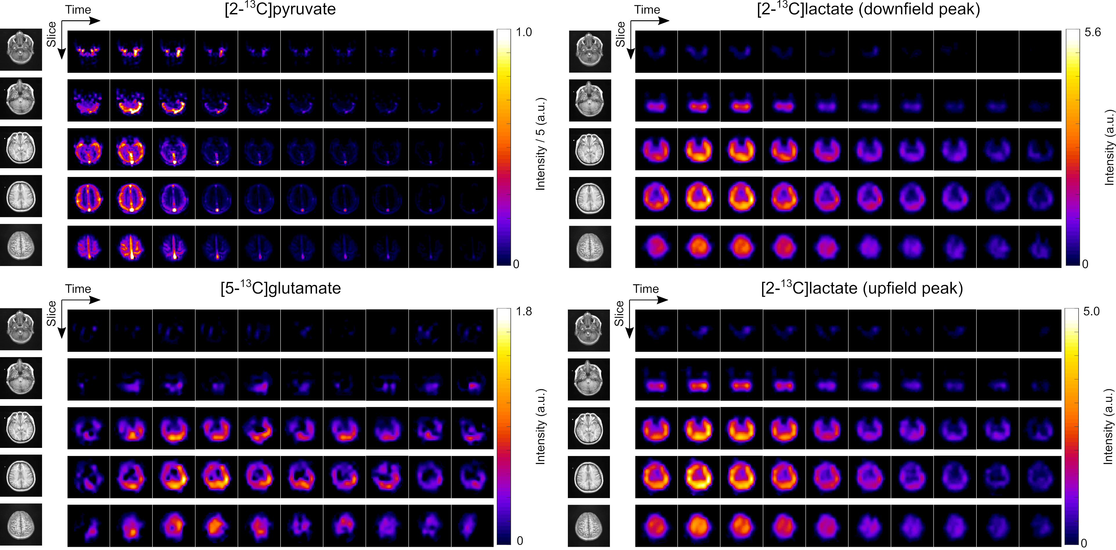
Figure 1. The 4D dynamics of hyperpolarized [2-13C]pyruvate, [5-13C]glutamate, [2-13C]lactate (downfield peak), and [2-13C]lactate (upfield peak) in the healthy human brain. The displayed images were obtained after denoising using a patch-based denoising method. The first 10 timeframes, acquired with a temporal resolution of 3 s, are displayed. The upper window level was adjusted to 20% of the maximum intensity voxel for pyruvate and to 100% for lactate and glutamate.
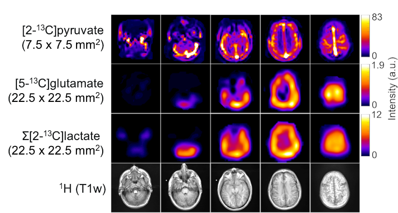
Figure 3. Representative AUC images of [2-13C]pyruvate, [5-13C]glutamate, and Σ[2-13C]lactate signals summed over 20 timeframes with 1H anatomical images from a healthy brain volunteer.