Hannes Michel Wiesner1, Rong Guo2,3, Yudu Li2,3, Yibo Zhao2,3, Zhi-Pei Liang2,3, Xiao-Hong Zhu1, and Wei Chen1
1CMRR, Department of Radiology, University of Minnesota, Minneapolis, MN, United States, 2Beckman Institute for Advanced Science and Technology, University of Illinois at Urbana-Champaign, Urbana, IL, United States, 3Departments of Electrical and Computer Engineering, University of Illinois at Urbana-Champaign, Urbana, IL, United States
1CMRR, Department of Radiology, University of Minnesota, Minneapolis, MN, United States, 2Beckman Institute for Advanced Science and Technology, University of Illinois at Urbana-Champaign, Urbana, IL, United States, 3Departments of Electrical and Computer Engineering, University of Illinois at Urbana-Champaign, Urbana, IL, United States
We demonstrated the feasibility and potential of the SPICE approach for obtaining high-fidelity 3D 31P MRSI of the human brain at 7T.
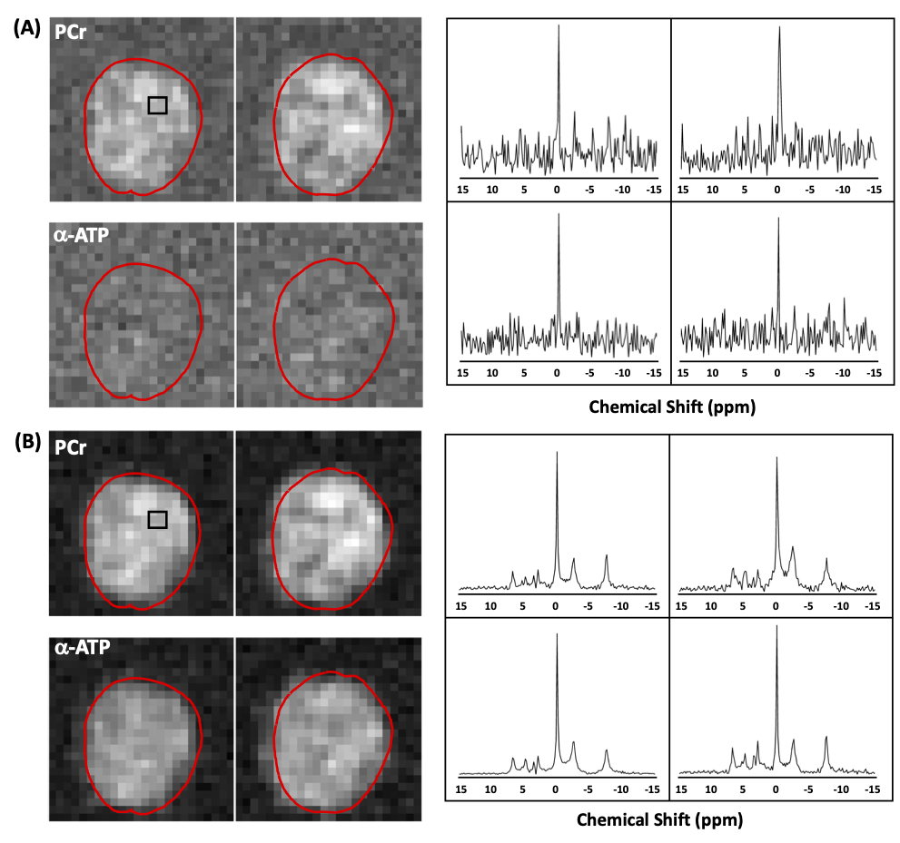
Figure 2 Representative in vivo human brain 31P CSI results from 3D dataset before (A) and after (B) using the SPICE denoising method. The data were acquired using Siemens CSI sequence with the following parameters: TR = 300 ms; TE = 0.35 ms; FOV = 220´220´200; Matrix = 24×24×12; Resolution (nominal) = 9.1×9.1×16 mm3; Average (weighted) = 10; bandwidth = 5000 Hz; vector size = 1024; scan time = 32 min. The red lines outline the brain edge and the left panels show the representative in vivo 31P spectra taken from 4 voxels within the black box as shown on the left panels.
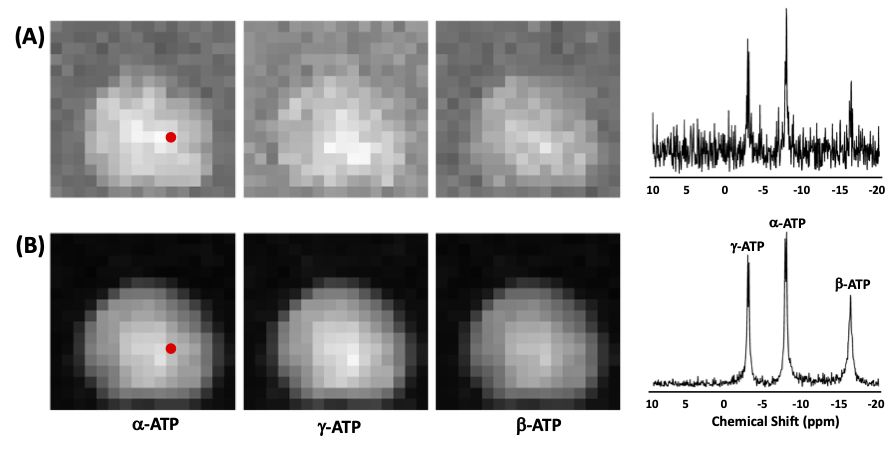
Figure 1 Representative phantom 31P CSI results from 3D dataset before (A) and after (B) using the SPICE denoising method and Siemens CSI sequence and parameters: TR = 200 ms; TE = 1.6 ms; FOV = 240×240×80; Matrix = 12×12×4; Resolution = 20×20×20 mm3; Average = 4; bandwidth = 5000 Hz; vector size = 800; scan time = 8 min.