Solène Bardin1,2, Michele Lecis1,3, Davide Boido1,2, Fawzi Boumezbeur1,2, and Luisa Ciobanu1,2
1NeuroSpin, CEA, Gif-sur-Yvette, France, 2Paris-Saclay University, Saclay, France, 3Technical University of Munich, Munich, Germany
1NeuroSpin, CEA, Gif-sur-Yvette, France, 2Paris-Saclay University, Saclay, France, 3Technical University of Munich, Munich, Germany
Accelerated CEST acquisitions using a linescan sequence coupled with an ultra-high magnetic field allows, for the first time, the detection of carnosine in vivo.
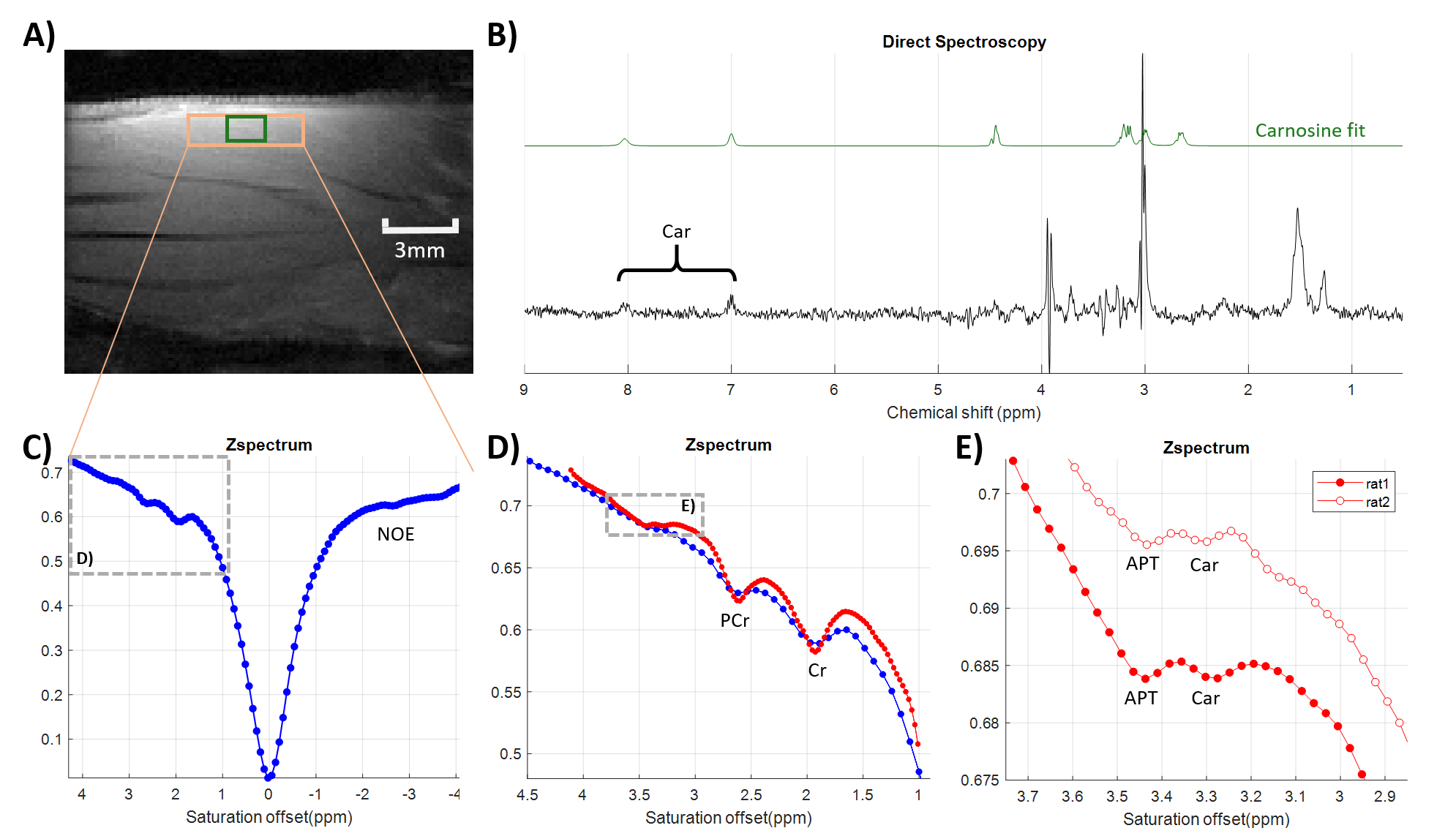
Figure 3. (A) Anatomical image of the lower-leg
muscle showing the voxels used for linescan and 1H-MRS (B) LASER spectrum (TE/TR=25/2000ms, 3*1.8*1.5mm3)
and the carnosine signal fitted using LCModel (C) Low resolution Z-spectrum (D)
Low (blue) and high (red) resolution Z-spectra showing Cr / PCr peaks (E) Zoom showing the Car and APT peaks
from two different rats. All peaks were detected with a significance p-value
< 0.001, except for the APT peak for rat 2 (p = 0.017). Full circles: rat 1,
Empty circles: rat 2.
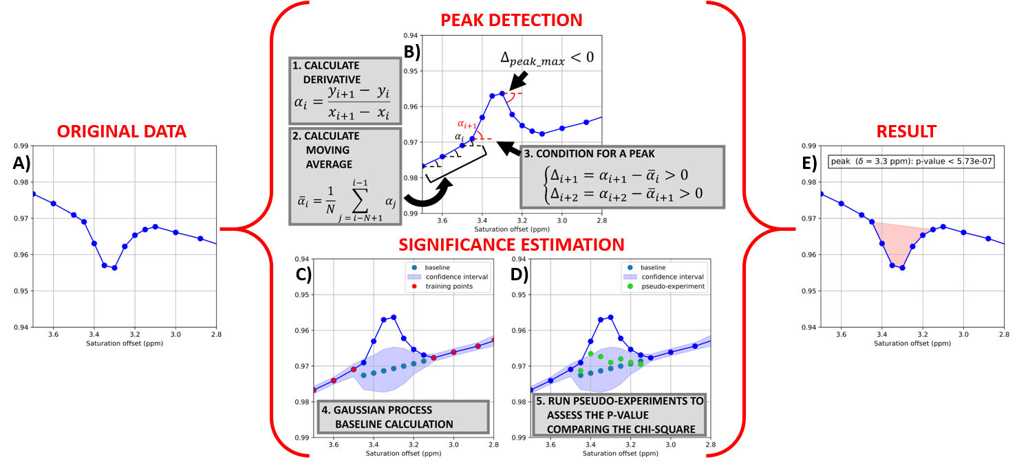
Figure 1. The “peak finder” is an algorithm that
automatically detects peaks and assesses their significance. The original data
(A) are flipped for peak detection (B). A baseline
is
estimated for each peak using Gaussian Processes
(GP)
and
a chi-square value χ2data is
calculated (C). The code randomly generates pseudo-experiments assuming a
Gaussian distribution around the baseline
with
a variance in accordance with the confidence interval returned by the GP (D).
The fraction of simulated curves with χ2pseudo < χ2data is
taken as p-value of the detected peak. The output is shown in (E).