Anna K. Scheipers1, Johanna Lott1, Armin M. Nagel1,2, Peter Bachert1, Mark E. Ladd1, and Tanja Platt1
1Medical Physics in Radiology, German Cancer Research Center (DKFZ), Heidelberg, Germany, 2University Hospital Erlangen, Institute of Radiology, Friedrich‐Alexander‐Universität Erlangen‐Nürnberg (FAU), Erlangen, Germany
1Medical Physics in Radiology, German Cancer Research Center (DKFZ), Heidelberg, Germany, 2University Hospital Erlangen, Institute of Radiology, Friedrich‐Alexander‐Universität Erlangen‐Nürnberg (FAU), Erlangen, Germany
Different corrections were applied to 23Na MR images of a phantom and of the human kidney and were invested regarding their influence on a reliable
quantification of the sodium concentration. Motion sorted B1+ correction was shown to be feasible with the applied setup for 23Na MRI at 7T.
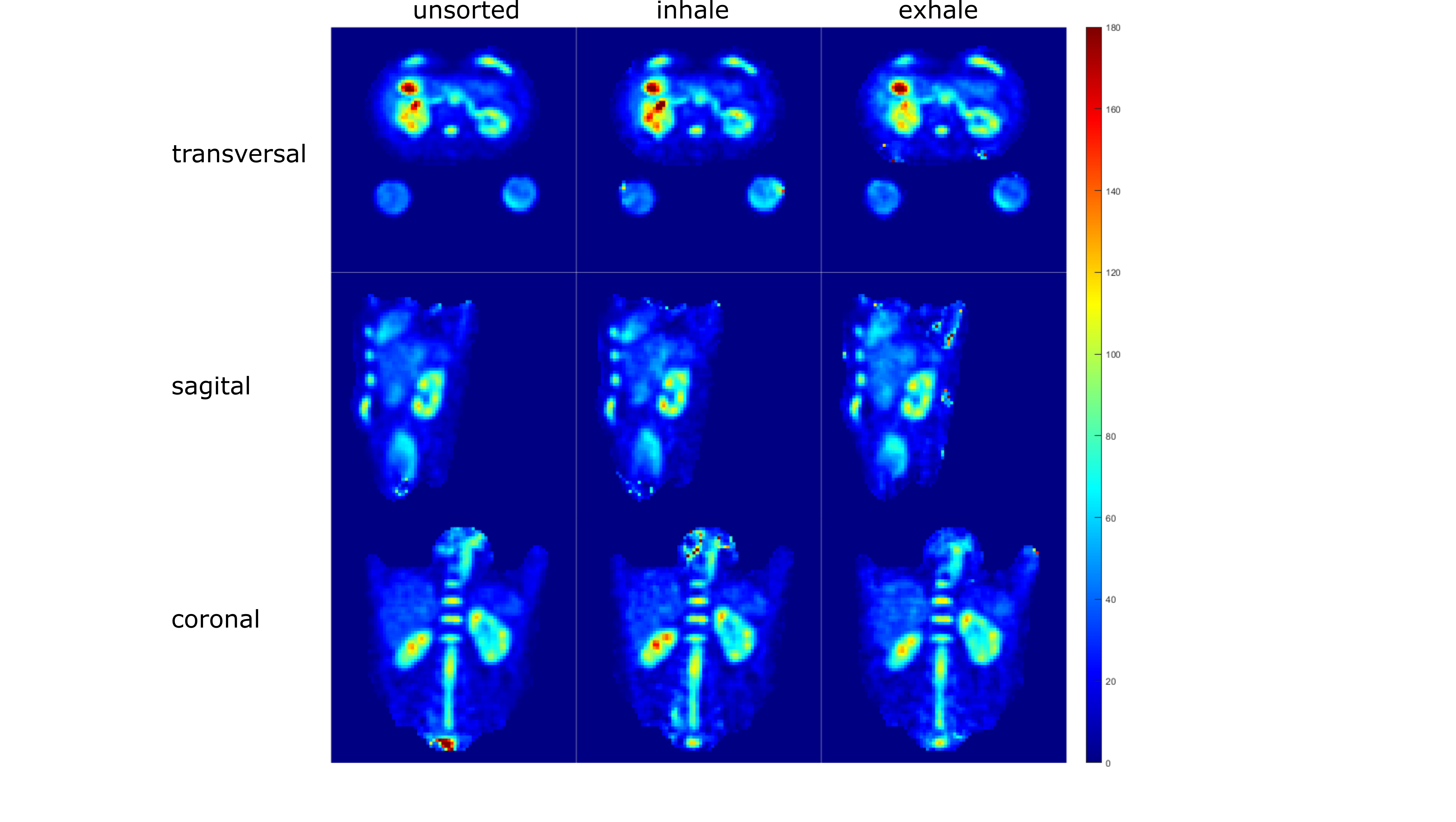
Fig. 3: Quantitative 23Na concentration in the human kidney at 7T in all directions for not respiratory sorted data, inhaled and exhaled state. Slices in each direction were chosen to have the kidneys visible (transversal: 38, coronal: 53, saggital: 33, for a 80×80×80 matrix).
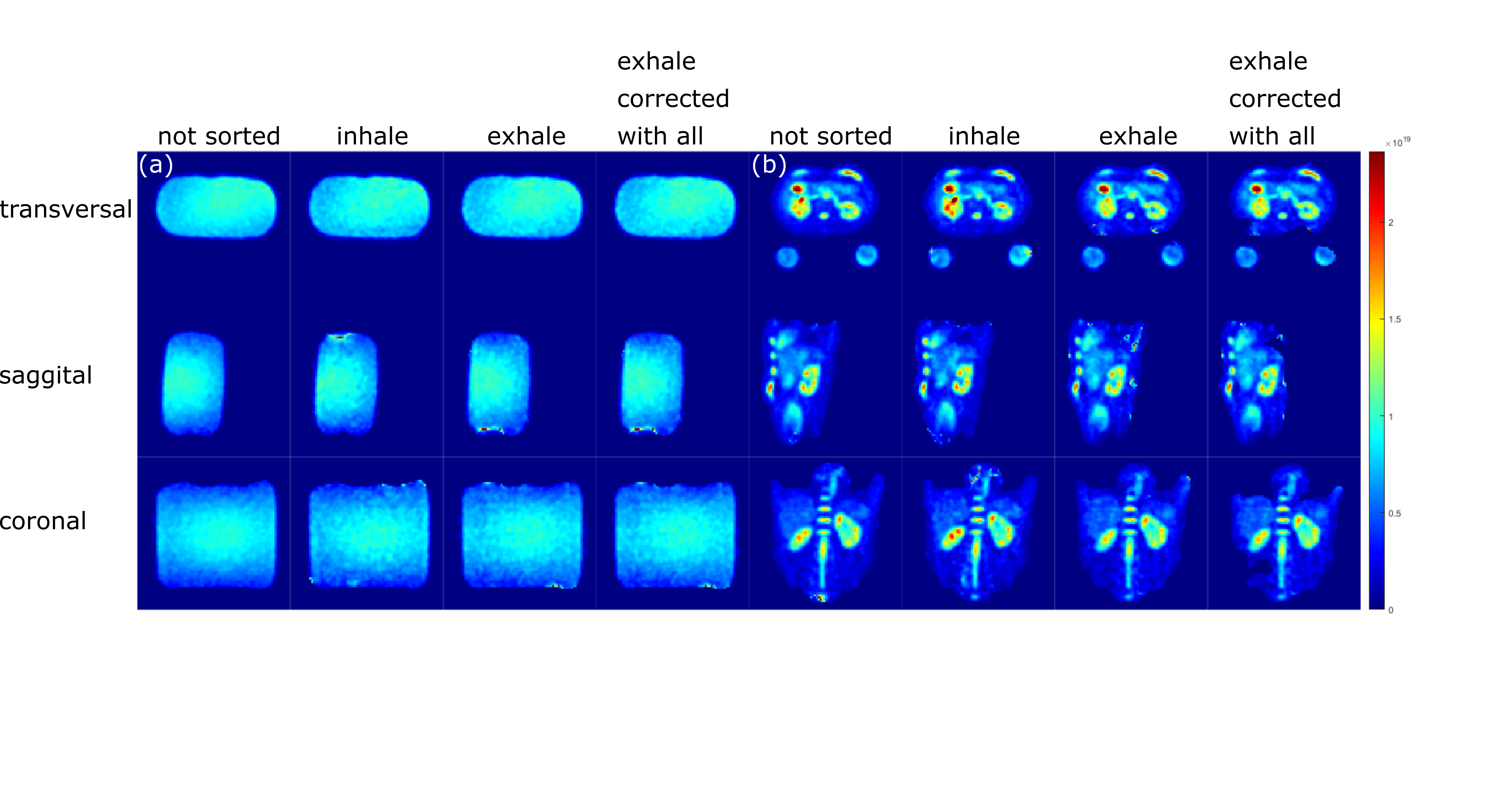
Fig. 2: Phantom (a) and in-vivo (b) data in all directions. First column shows the not respiratory sorted data corrected with the unsorted B1+ map, second column shows the inhaled state data corrected with the inhale sorted B1+ map, third and fourth column show the exhaled state data corrected with the exhale sorted B1+ map and the unsorted B1+ map respectively. Slices in each direction were chosen to have the kidneys visible (transversal: 38, coronal: 53, saggital: 33, for a 80×80×80 matrix).