Zidan Yu1,2, Olga Dergahyova1, Daniel K. Sodickson1,2, Guillaume Madelin1,2, and Martijn A. Cloos3,4
1Center for Biomedical Imaging, Department of Radiology, New York University School of Medicine, New York, NY, United States, 2Vilcek Institute of Graduate Biomedical Sciences, NYU Langone Health, New York, NY, United States, 3Centre for Advanced Imaging, The University of Queensland, Brisbane, Australia, 4ARC Training Centre for Innovation in Biomedical Imaging Technology, The University of Queensland, Brisbane, Australia
1Center for Biomedical Imaging, Department of Radiology, New York University School of Medicine, New York, NY, United States, 2Vilcek Institute of Graduate Biomedical Sciences, NYU Langone Health, New York, NY, United States, 3Centre for Advanced Imaging, The University of Queensland, Brisbane, Australia, 4ARC Training Centre for Innovation in Biomedical Imaging Technology, The University of Queensland, Brisbane, Australia
In this work, we present an optimized 3D technique that can simultaneously acquire quantitative 1H density, T1, T2, B1+ maps and a 23Na image of the whole head in a reasonable scan time (~20 min).
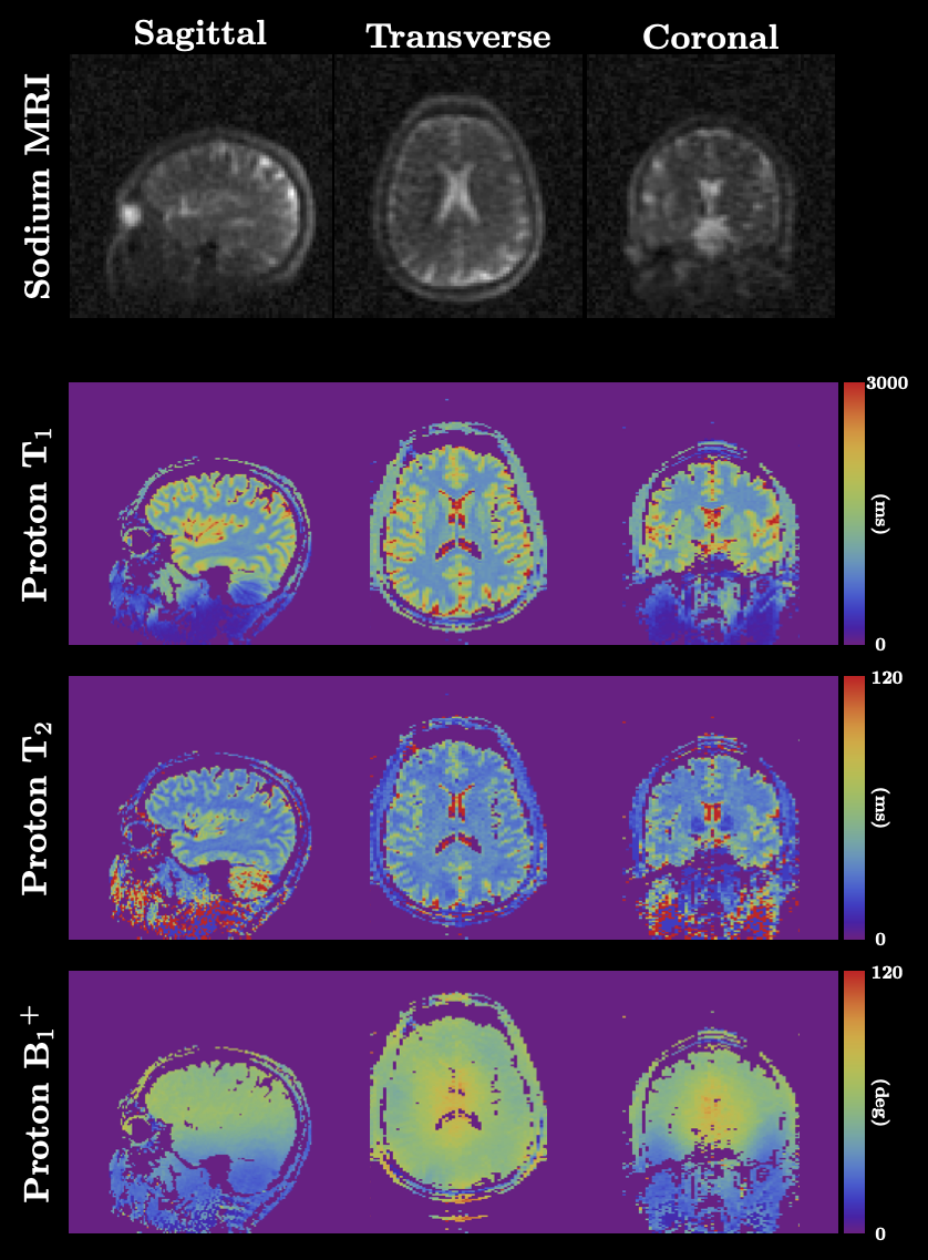
Figure 4: In-vivo results. Sodium images are shown in the first row, which has 3 mm3 isotropic resolution. Proton T1,T2, B1+ maps from the three directions are shown beneath the sodium images.
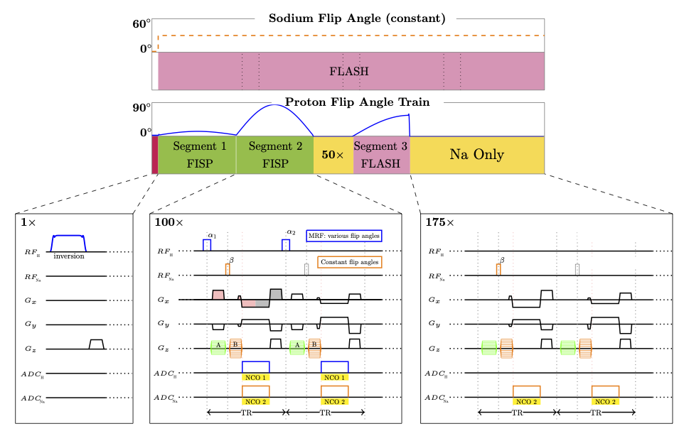
Figure 1: Sequence diagram of the 3D simultaneous technique. The bars on top show the sodium excitations with constant flip angle and the proton MRF train with various flip angles. The details of the sequence for different segments are shown in the corresponding cards on the bottom.