Dana C. Peters1, Stefan Markovic2, Qingjia Bao2, Dina Preise2, Keren Sasson2, Lilach Agemy2, Avigdor Scherz2, and Lucio Frydman2
1Radiology and Biomed Eng., Yale University, New Haven, CT, United States, 2Weizmann Institute of Science, Rehovot, Israel
1Radiology and Biomed Eng., Yale University, New Haven, CT, United States, 2Weizmann Institute of Science, Rehovot, Israel
Multi-echo bSSFP increased the SNR for Deuterium Metabolic Imaging (DMI), and was able to spectrally resolve glucose, water and lactate.
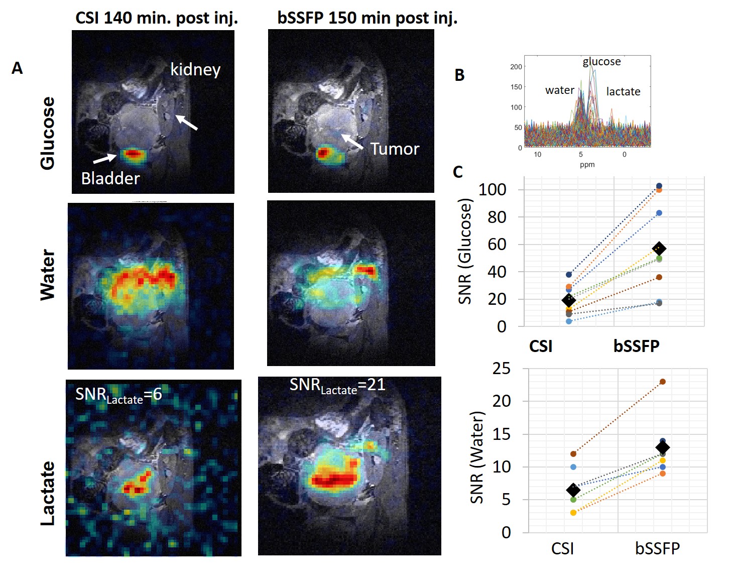
Figure 5: Comparing
32x32 ME-bSSFP and CSI data arising from a large pancreatic
tumor in a mouse at ~2 hrs after injection of deuterated glucose. The regional distribution of the metabolites
is similar (color, superimposed on 1H anatomical images in
grayscale), with glucose located in the bladder, lactate in part of the
tumor, and water diffusely distributed. (B) CSI data
for all pixels, with lactate visible on the spectrum. C) SNR comparisons for matched ROIs. Glucose SNR was tripled with bSSFP vs. CSI (57 ±30 vs. 19±11,
p<0.001, N=10). Water SNR was doubled
(13±5 vs. 7 ±3, p=0.005, N=8).

Figure 4: Phantom study with 3 tubes containing deuterated water, glucose
and lactate. (A) ME-bSSFP isolation of the different metabolites, with some
cross-talk. (B) 2H NMR spectra summarizing the CSI data for all
spatial elements, showing the different metabolites. (C) Summary of the SNR
measurements showing significant improvements for lactate and glucose. The “avg bSSFP SNR” was estimated as the SNR
obtained by simple signal averaging over the metabolite’s region (i.e., with no
IDEAL processing).