Sophie Queler1, Ek Tsoon Tan1, Martin Prince2, John Carrino1, and Darryl Sneag1
1Radiology and Imaging, Hospital for Special Surgery, New York, NY, United States, 2Weill Cornell Medicine, New York, NY, United States
1Radiology and Imaging, Hospital for Special Surgery, New York, NY, United States, 2Weill Cornell Medicine, New York, NY, United States
Ferumoxytol, an iron oxide nanoparticle, improves vascular suppression in magnetic resonance neurography of the brachial plexus compared to non-contrast techniques.
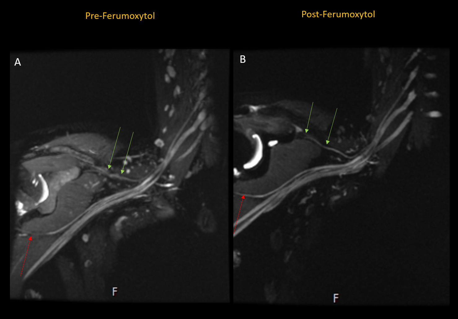
Figure 2. Pre-ferumoxytol 3D
STIR-FSE multiplanar
reformatted (MPR) maximal intensity projection (MIP) (a) demonstrates venous
contamination obscuring the suprascapular (green arrows) and axillary (red arrow)
nerves. On the post-ferumoxytol MPR MIP (b) the suprascapular (green arrows) and axillary (red arrow)
nerves are clearly delineated.
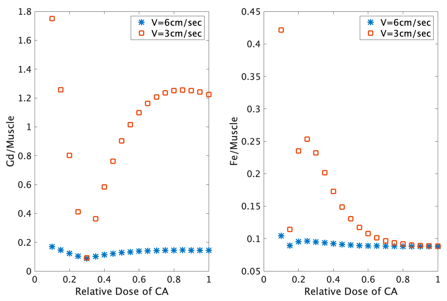
Figure 1. Simulations of different relative doses
of contrast agents (CA), for (a) Gadolinium+blood to
muscle contrast, and (b) Ferumoxytol+blood to
muscle contrast, at two different blood velocities, assuming TE=80 ms and
TI=255 ms.