Christof Boehm1, Marianne Goeger-Neff2, Hendrik T. Mulder3, Benjamin Zilles2, Lars H. Lindner2, Gerard C. van Rhoon3, Dimitrios C. Karampinos4, and Mingming Wu1
1Technical University of Munich, Munich, Germany, 2Department of Medicine III, University Hospital, LMU Munich, Munich, Germany, 3Erasmus MC Cancer Institute, Rotterdam, Netherlands, 4Department of Diagnostic and Interventional Radiology, School of Medicine, Technical University of Munich, Munich, Germany
1Technical University of Munich, Munich, Germany, 2Department of Medicine III, University Hospital, LMU Munich, Munich, Germany, 3Erasmus MC Cancer Institute, Rotterdam, Netherlands, 4Department of Diagnostic and Interventional Radiology, School of Medicine, Technical University of Munich, Munich, Germany
MR thermometry monitoring of mild RF hyperthermia of tumors in the pelvis and the upper leg are hampered by susceptibility artefacts. Novel methods from quantitative susceptibility mapping such as total field inversion resolve susceptibility artefacts while preserving temperature.
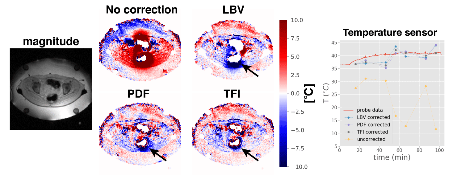
Figure 4: Resulting
temperature maps for a cervical cancer patient during mild RF-HT of the tumor.
Particularly for tumors close to the intestines, gas motion causes severe
artifacts. The black arrows point at residual phase errors after background
field correction, that appears to be less in the TFI. Furthermore, the LBV
method resulted in the loss of valuable pixels. The comparison of the corrected
temperature with a sensor illustrates how severe the susceptibility artifacts were
in the uncorrected DEGRE.
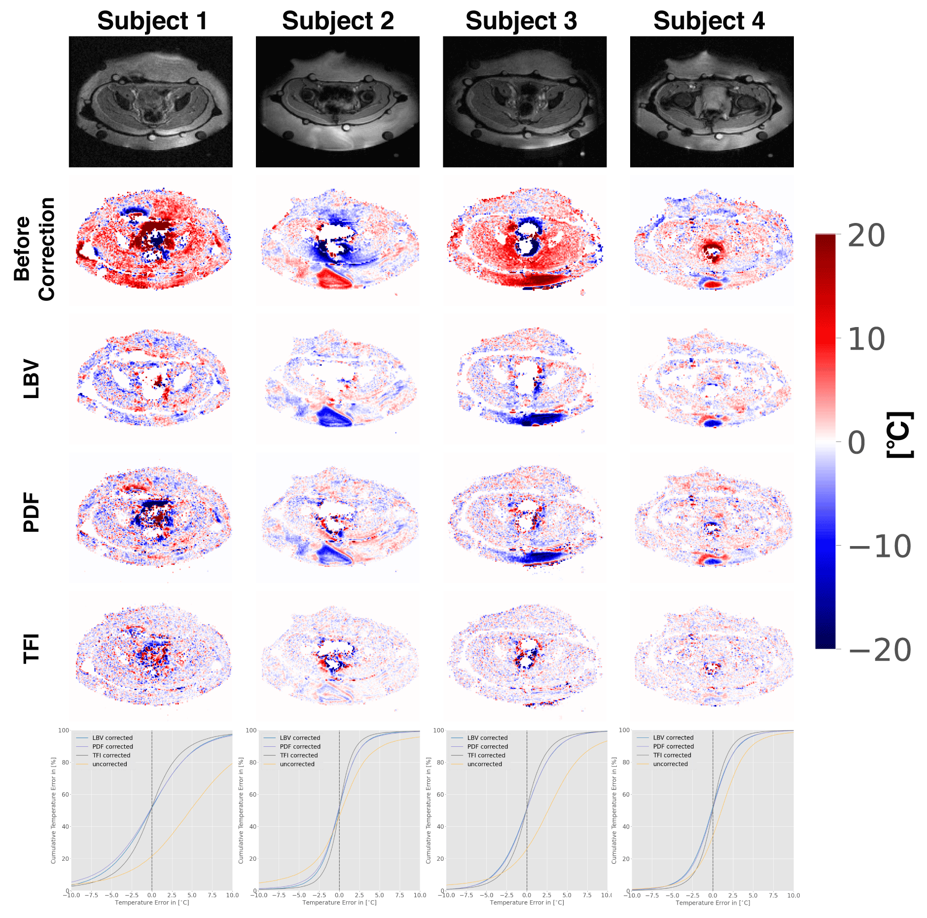
Figure 3: Bowel
motion-induced susceptibility artefacts in volunteers at constant temperature
and their correction with LBV, PDF and TFI. The temperature error maps before
and after correction are displayed.
In
the cumulative error plots, the ratio is calculated between the numbers of voxels occupying the given value
and less over all voxel counts.
In contrast to the simulation results, as
seen in Fig.1, the cumulative error plots (last row) indicate that TFI results
in the least residual phase errors.