Kisoo Kim1, Chris Diederich2, and Eugene Ozhinsky1
1Department of Radiology & Biomedical Imaging, University of California, San Francisco, San Francisco, CA, United States, 2Department of Radiation Oncology, University of California, San Francisco, San Francisco, CA, United States
1Department of Radiology & Biomedical Imaging, University of California, San Francisco, San Francisco, CA, United States, 2Department of Radiation Oncology, University of California, San Francisco, San Francisco, CA, United States
Motion-robust, multi-slice, real-time MR thermometry reconstruction pipeline was developed for accurate and stable temperature measurements in abdominal organs.
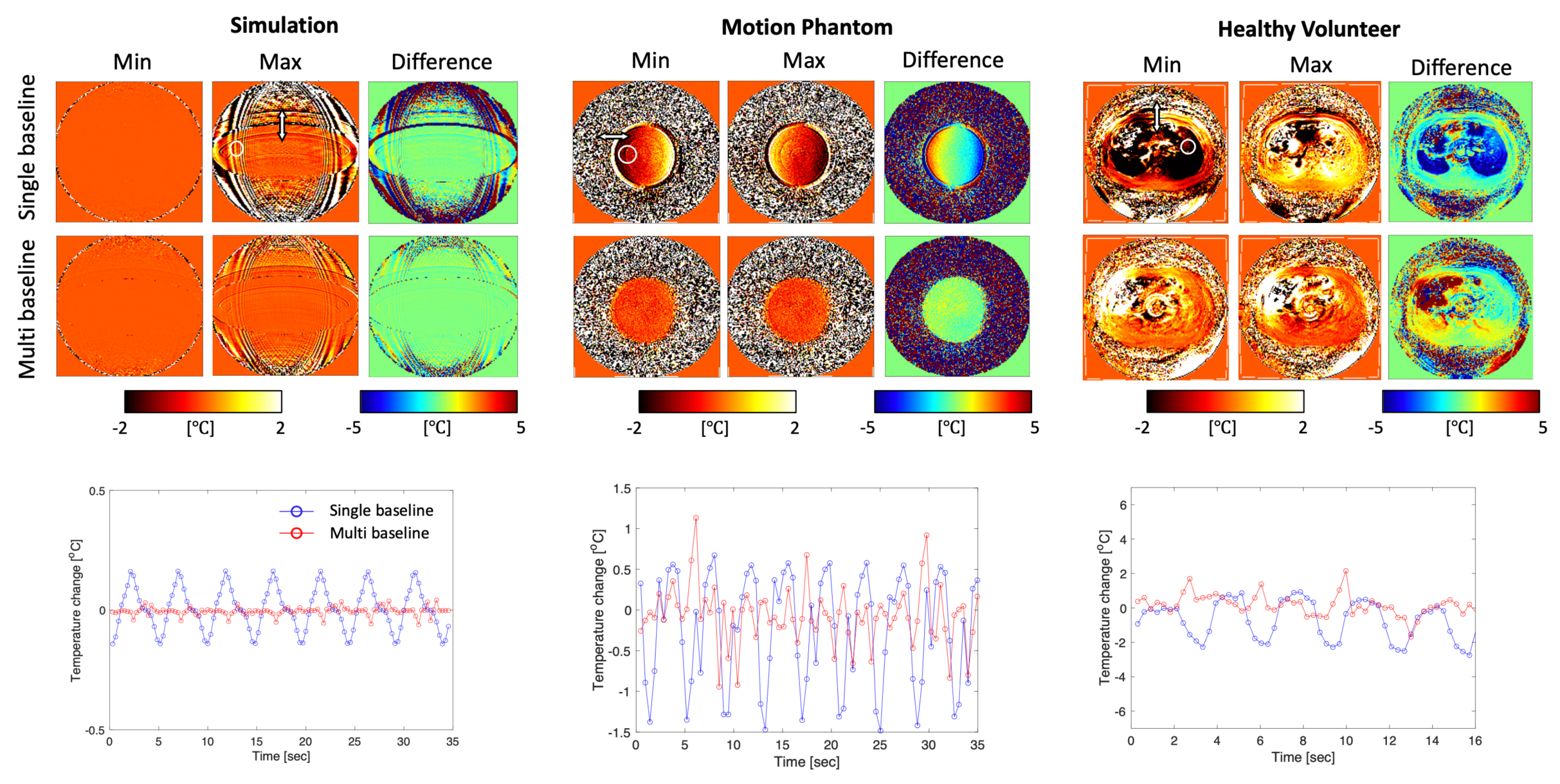
Figure 3. Single- and multi-baseline PRFS temperature maps at two positions of the respiratory cycle and plots of temperature change within an ROI (white circle) in simulation, phantom experiment with respiratory motion simulator, and in free-breathing acquisition in a healthy volunteer without heating. Temperature difference maps between two positions of the cycle (max-min) show that the multi-baseline reconstruction results in less motion artifacts compared to the single baseline reconstruction throughout the breathing cycle.
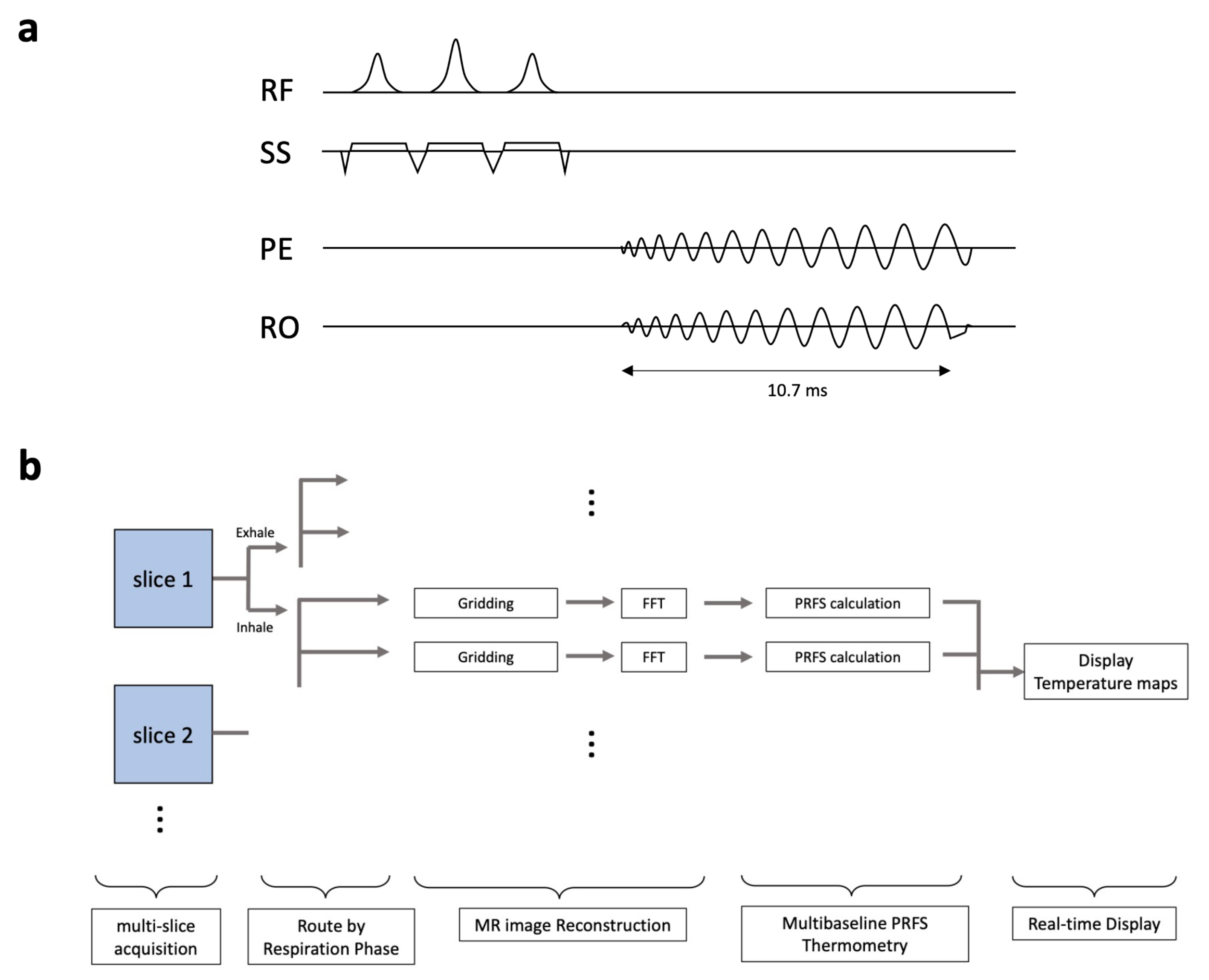
Figure 1. (a) schematic diagram of spiral-based MR GRE sequence and (b) real-time reconstruction pipeline. RF: RF pulse excitation, SS: Slice-Selection gradient, PE: Phase Encoding gradient, RO: Read-Out gradient. FFT: Fast Fourier Transform.