Ann S Choe1,2,3, Bohao Tang4, Kimberly R. Smith5, Hamed Honari6, Martin A. Lindquist4, Brian S. Caffo4, and James J. Pekar1,3
1F.M. Kirby Research Center for Functional Brain Imaging, Kennedy Krieger Institute, Baltimore, MD, United States, 2International Center for Spinal Cord Injury, Kennedy Krieger Institute, Baltimore, MD, United States, 3Department of Radiology, Johns Hopkins Medicine, Baltimore, MD, United States, 4Department of Biostatistics, Johns Hopkins Bloomberg School of Public Health, Baltimore, MD, United States, 5Department of Psychiatry and Behavioral Sciences, Johns Hopkins Medicine, Baltimore, MD, United States, 6Department of Electrical and Computer Engineering, Johns Hopkins University, Baltimore, MD, United States
1F.M. Kirby Research Center for Functional Brain Imaging, Kennedy Krieger Institute, Baltimore, MD, United States, 2International Center for Spinal Cord Injury, Kennedy Krieger Institute, Baltimore, MD, United States, 3Department of Radiology, Johns Hopkins Medicine, Baltimore, MD, United States, 4Department of Biostatistics, Johns Hopkins Bloomberg School of Public Health, Baltimore, MD, United States, 5Department of Psychiatry and Behavioral Sciences, Johns Hopkins Medicine, Baltimore, MD, United States, 6Department of Electrical and Computer Engineering, Johns Hopkins University, Baltimore, MD, United States
Using concurrent cutaneous electrogastrography and resting-state fMRI in a highly-sampled individual experimental design, a majority of resting-state brain networks were found to be phase synchronized with the intrinsic rhythm of the stomach.
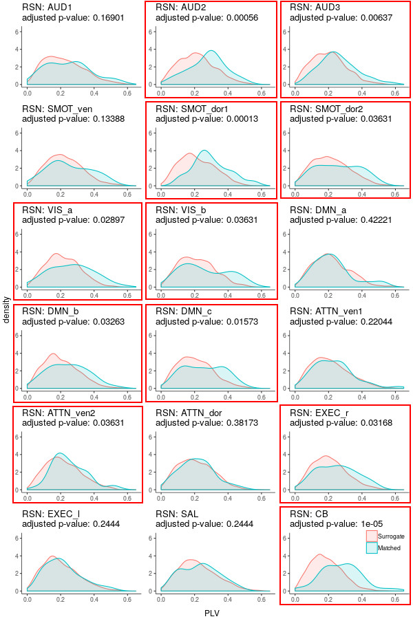
Fig 3. Gaussian kernel density estimates (i.e., smoothed histograms) of awPLV between resting-state networks and gastric signals recorded concurrently (cyan) and on different days (coral). P-values from Wilcoxon rank tests have been adjusted for multiple comparisons using a FDR of 0.05. Subfigures for networks with significant gastric synchronization are outlined in red.
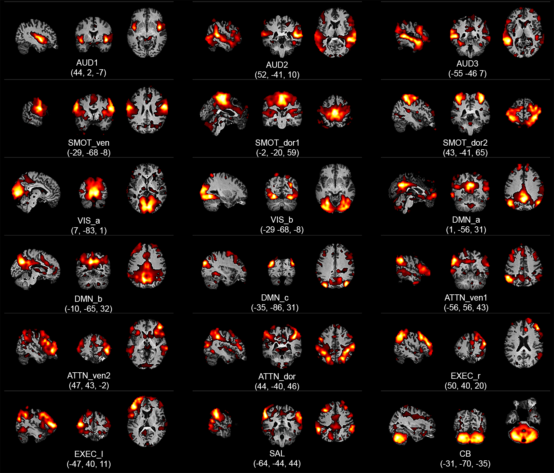
Fig 2. Aggregate spatial maps of resting-state networks (RSNs). Representative sagittal, coronal, and axial views (left-to-right) are overlaid on structural images in the Montreal Neurological Institute template space. Coordinates (in mm) for each view are given below each subfigure. (AUD: auditory, SMOT: somatosensory-motor, VIS: visual, DMN: default mode network, ATTN: attention, EXEC: executive, SAL: salience, CB: cerebellar, ven: ventral, dor: dorsal, r: right, l: left). Figure adapted from 1.