Jitka Starekova1, Lloyd Estkowski2, Ruiqi Geng1,3, Yuxin Zhang1,3, Diego Hernando1,3, and Scott B Reeder1,3,4,5,6
1Radiology, University of Wisconsin-Madison, Madison, WI, United States, 2Global MR Applications and Workflow GE Healthcare, Madison, WI, United States, 3Medical Physics, University of Wisconsin-Madison, Madison, WI, United States, 4Biomedical Engineering, University of Wisconsin-Madison, Madison, WI, United States, 5Medicine, University of Wisconsin-Madison, Madison, WI, United States, 6Emergency Medicine, University of Wisconsin-Madison, Madison, WI, United States
1Radiology, University of Wisconsin-Madison, Madison, WI, United States, 2Global MR Applications and Workflow GE Healthcare, Madison, WI, United States, 3Medical Physics, University of Wisconsin-Madison, Madison, WI, United States, 4Biomedical Engineering, University of Wisconsin-Madison, Madison, WI, United States, 5Medicine, University of Wisconsin-Madison, Madison, WI, United States, 6Emergency Medicine, University of Wisconsin-Madison, Madison, WI, United States
This prospective study optimized an esophageal MRI protocol including high-resolution T2-weighted imaging with deep learning denoising, as well as motion-robust diffusion-weighted imaging (DWI).
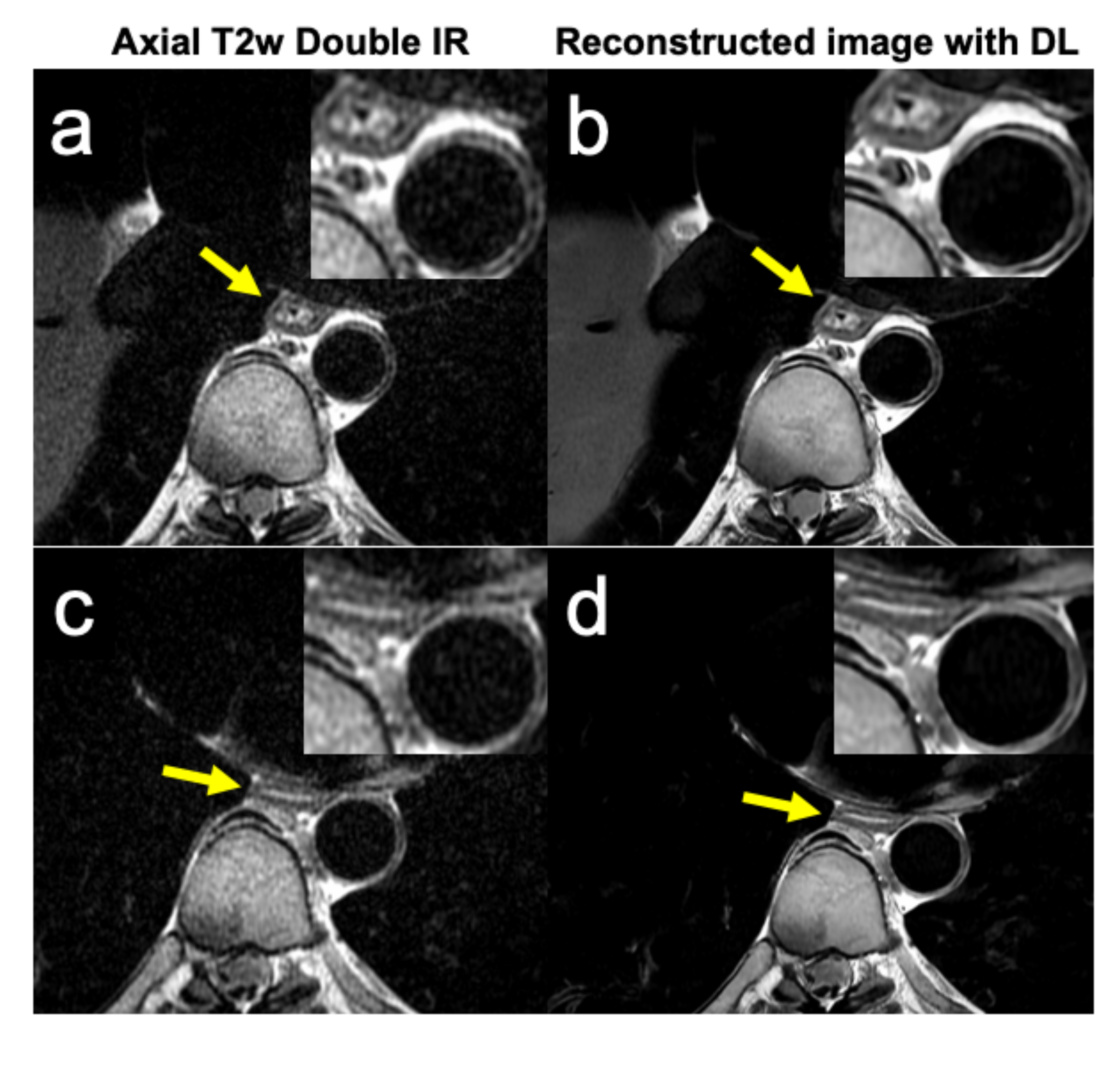
Figure 3. High-resolution T2w imaging of esophagus (a and c) is further improved using DL reconstruction with commercially available AIR Recon DL software (GE Healthcare, Waukesha, WI)10. Shown are images of distal esophagus of a 37-year-old male healthy volunteer. Abbreviations: T2w double IR, T2 weighted double inversion recovery; DL, deep learning
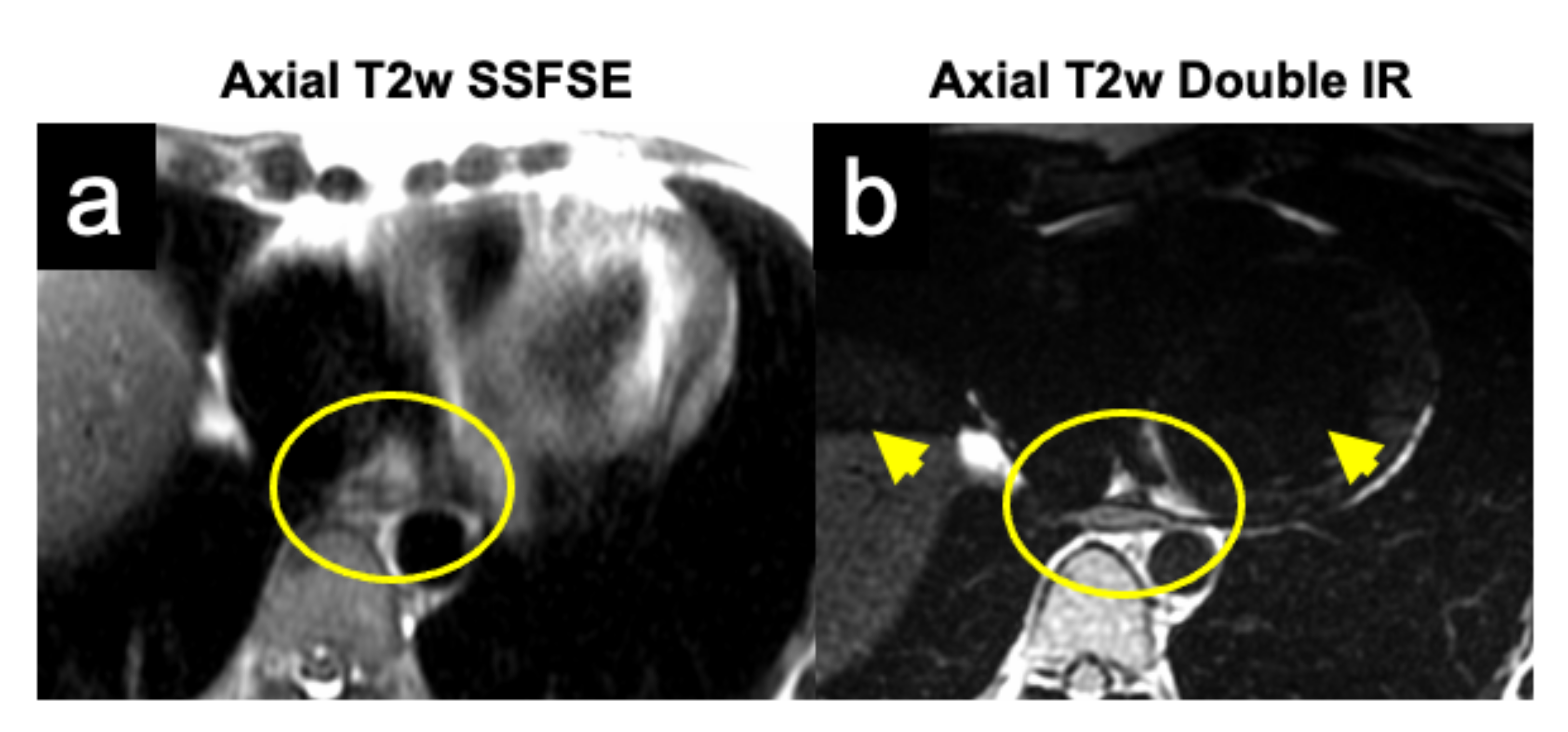
Figure 2. Cardiac induced motion artifacts diminish image quality of T2w image of esophagus (esophagus is in the center of yellow circles) and may mimic lesion in esophageal wall. Cardiac gating and saturation bands (arrows) help to overcome this limitations. Abbreviations: T2w SSFSE, T2 weighted single shot fast spin echo; T2w double IR, T2 weighted double inversion recovery