Yufei Zhao1,2, Yang Jiang1,2, Jingyue Dai1,2, Honghong Wu1,2, Ying Cui1,2, Xinxiang Li1,2, and Xingui Peng1,2
1Jiangsu Key Laboratory of Molecular and Functional Imaging, Southeast University, Nanjing, China, 2Radiology, Zhongda Hospital Southeast University, Nanjing, China
1Jiangsu Key Laboratory of Molecular and Functional Imaging, Southeast University, Nanjing, China, 2Radiology, Zhongda Hospital Southeast University, Nanjing, China
T1ρ magnetic resonance can non-invasively assess the changes in the distribution of myofiber in male T2DM patients and this changes is gradually obvious as the course of the disease progresses.
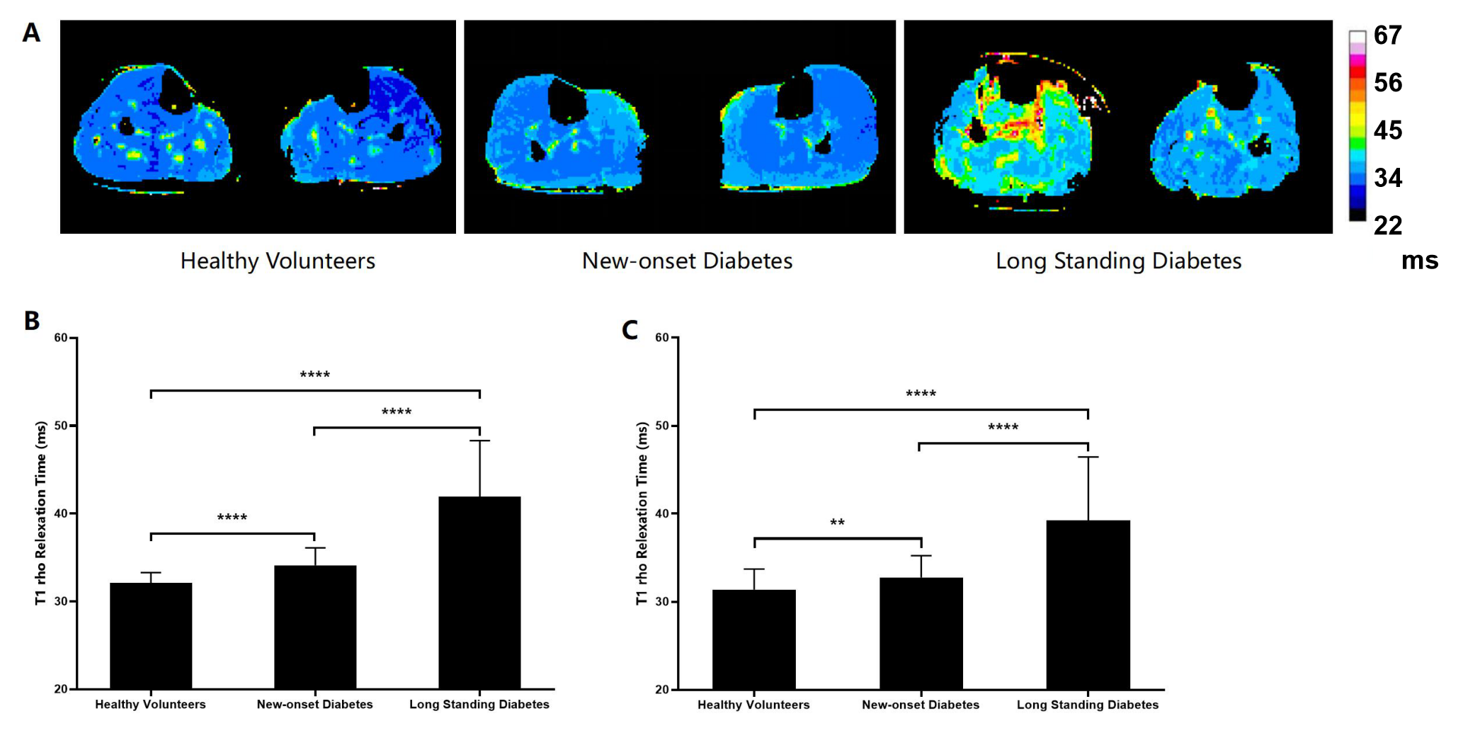
Comparison of the T1ρ relaxation time of the TA and SOL muscles in the three group. A) Comparison of the T1ρ map of the TA and SOL muscles in the three group. B) The T1ρ relaxation times of the SOL muscle in the three group. C) The T1ρ relaxation times of the TA in the three group.**P < 0.01 and ****P < 0.0001.
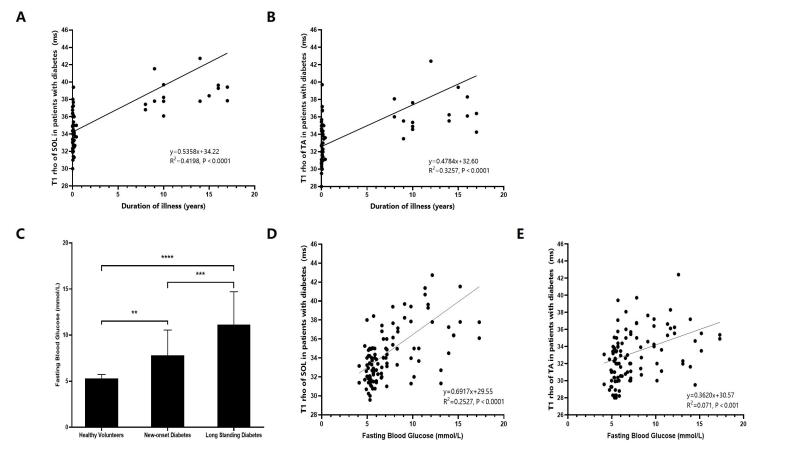
The linear relationship with the duration time of illness and fasting blood glucose of the TA and SOL muscles. A) The linear relationship with the duration time of illness of the TA muscles. B) The linear relationship with the duration time of illness of the SOL muscles. C) Fasting blood glucose levels in three groups. D) The linear relationship with the fasting blood glucose and the SOL muscles T1ρ relaxation time. E) The linear relationship with the fasting blood glucose and the TA muscles T1ρ relaxation time. **P < 0.01, ***P < 0.001 and ****P < 0.0001.