Jill T Shah1, Katherine Medina2,3, Haresh R Rajamohan2,4, Justin Ho2,3, Cem M Deniz2,3, and Ryan Brown2,3
1New York University Grossman School of Medicine, New York, NY, United States, 2Center for Biomedical Imaging, Department of Radiology, New York University Grossman School of Medicine, New York, NY, United States, 3Center for Advanced Imaging Innovation and Research (CAI2R), Department of Radiology, New York University Grossman School of Medicine, New York, NY, United States, 4Center for Data Science, New York University, New York, NY, United States
1New York University Grossman School of Medicine, New York, NY, United States, 2Center for Biomedical Imaging, Department of Radiology, New York University Grossman School of Medicine, New York, NY, United States, 3Center for Advanced Imaging Innovation and Research (CAI2R), Department of Radiology, New York University Grossman School of Medicine, New York, NY, United States, 4Center for Data Science, New York University, New York, NY, United States
We developed an automated algorithm based on a convolutional neural network to segment posterior calf muscles. We used the tool to show elevated fat fraction in gastroc medial and soleus muscles of individuals with diabetic peripheral neuropathy.
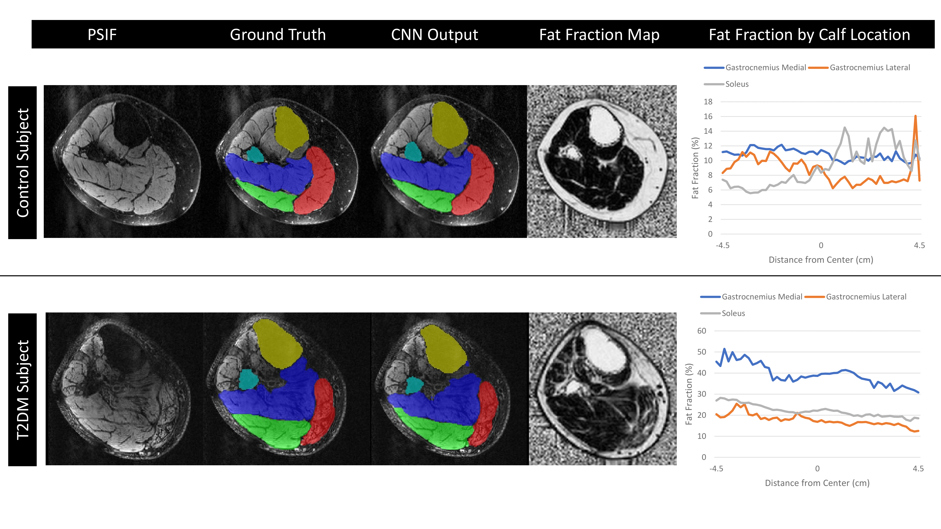
Figure 1. Results from one subject from the control cohort and a second subject from the T2DM cohort. The anatomic PSIF MR image of one slice is shown, as well as the corresponding ground truth segmentation and CNN segmentation results. The fat fraction map of the same slice is also shown. For each subject, a graph is depicted on the right, highlighting the variation in fat fraction as a function of slice location along the calf for the gastroc medial, gastroc lateral, and soleus muscles.
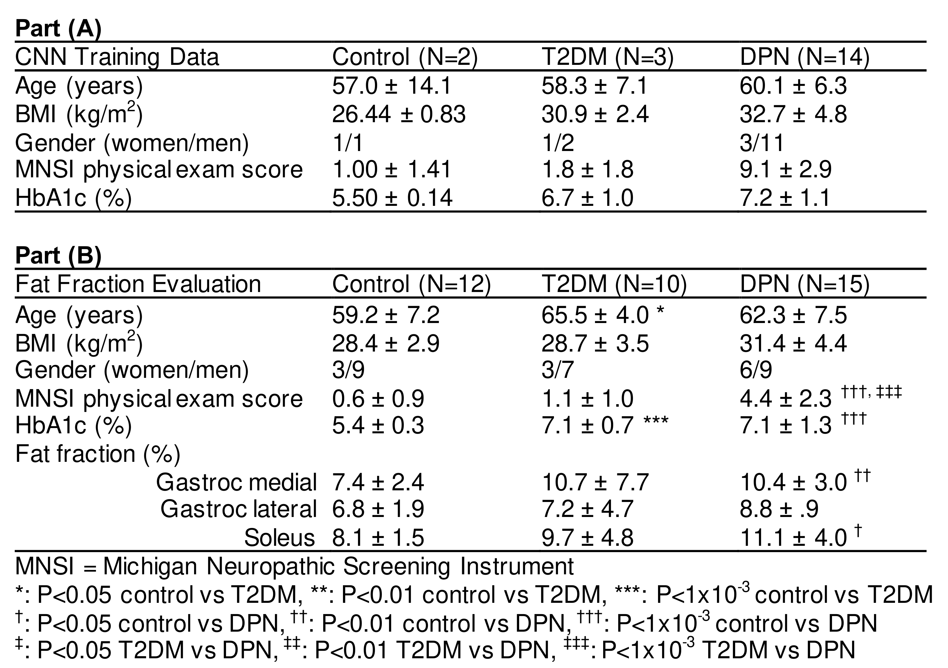
Table 1. Participant characteristics for the training (N=19) and fat fraction evaluation (N=37) cohorts. Fat fraction percentages were calculated over an 8.1cm longitudinal region using the automated segmentation tool.