Galina E Pavlovskaya1,2, Christopher J Philp1, Thomas Meersmann1, Petra Hanson3,4, Harpal S Randeva4,5, Paul Paul O’Hare4,5, and Thomas Barber4,5
1SPMIC/Medicine, Univeristy of Nottingham, Nottingham, United Kingdom, 24Nottingham NIHR Biomedical Research Centre, Nottingham, United Kingdom, 3Warwick Medical School, University of Warwick, Warwick, United Kingdom, 42Warwickshire Institute for the Study of Diabetes Endocrinology and Metabolism, University Hospitals Coventry and Warwickshire, Coventry, United Kingdom, 5Warwick Medical School, University of Warwick, Coventry, United Kingdom
1SPMIC/Medicine, Univeristy of Nottingham, Nottingham, United Kingdom, 24Nottingham NIHR Biomedical Research Centre, Nottingham, United Kingdom, 3Warwick Medical School, University of Warwick, Warwick, United Kingdom, 42Warwickshire Institute for the Study of Diabetes Endocrinology and Metabolism, University Hospitals Coventry and Warwickshire, Coventry, United Kingdom, 5Warwick Medical School, University of Warwick, Coventry, United Kingdom
Sodium skin storage capacity probed by ultra high field 23Na
MRS is diminished in Type 2 Diabetes Mellitus patients.
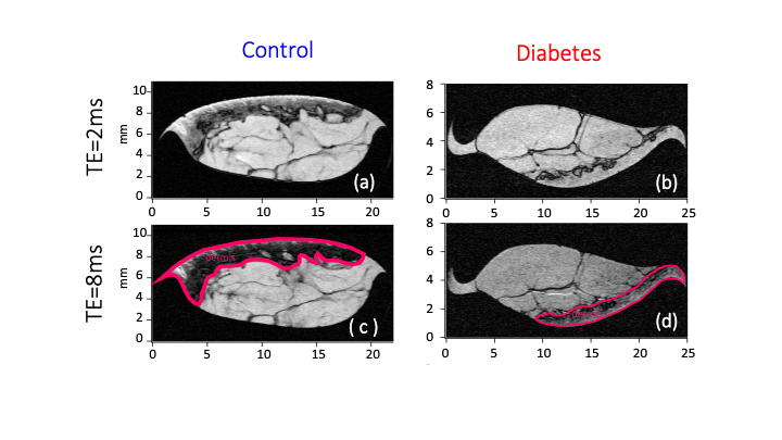
Figure 1. Anatomical compartments (a-b) of the human the
skin using 1H 9.4T MRI approaching histological length scales (20 mm) for control (NDB2) and diabetic (DB3) skin
biopsies. Segmentation of dermis layer in in control (c) and diabetic (d)
biopsies with the increase of TE to 8ms.
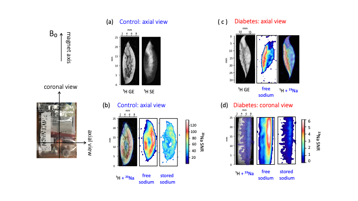
Figure 2. (a) - 1H high resolution
axial plane MRI of control (NDB8) skin biopsy using gradient echo MRI protocol
with TE = 2ms (1H GE). Enhanced segmentation of the dermis layer
(appears dark in 1H SE) was obtained with spin-echo MRI using the
same as in 1H GE geometry;
(b) – free sodium image of control biopsy; (c) - 1H GE of the diabetic skin biopsy DB3 in the axial
plane with corresponding free sodium 23Na MRI and co-registration
image; (d) - coronal projection DB3 biopsy. Free and
stored sodium spaces are co-registered within the dermis compartment.