Yu Veronica Sui1 and Mariana Lazar1
1Radiology, New York University Grossman School of Medicine, New York, NY, United States
1Radiology, New York University Grossman School of Medicine, New York, NY, United States
Using the nonlinearity index of cortical myelin profile, we observed substantial age-related microstructural changes in addition to those captured by macroscale cortical thickness measures. These results highlight layer-specific demyelination and neurodegeneration in normal aging.
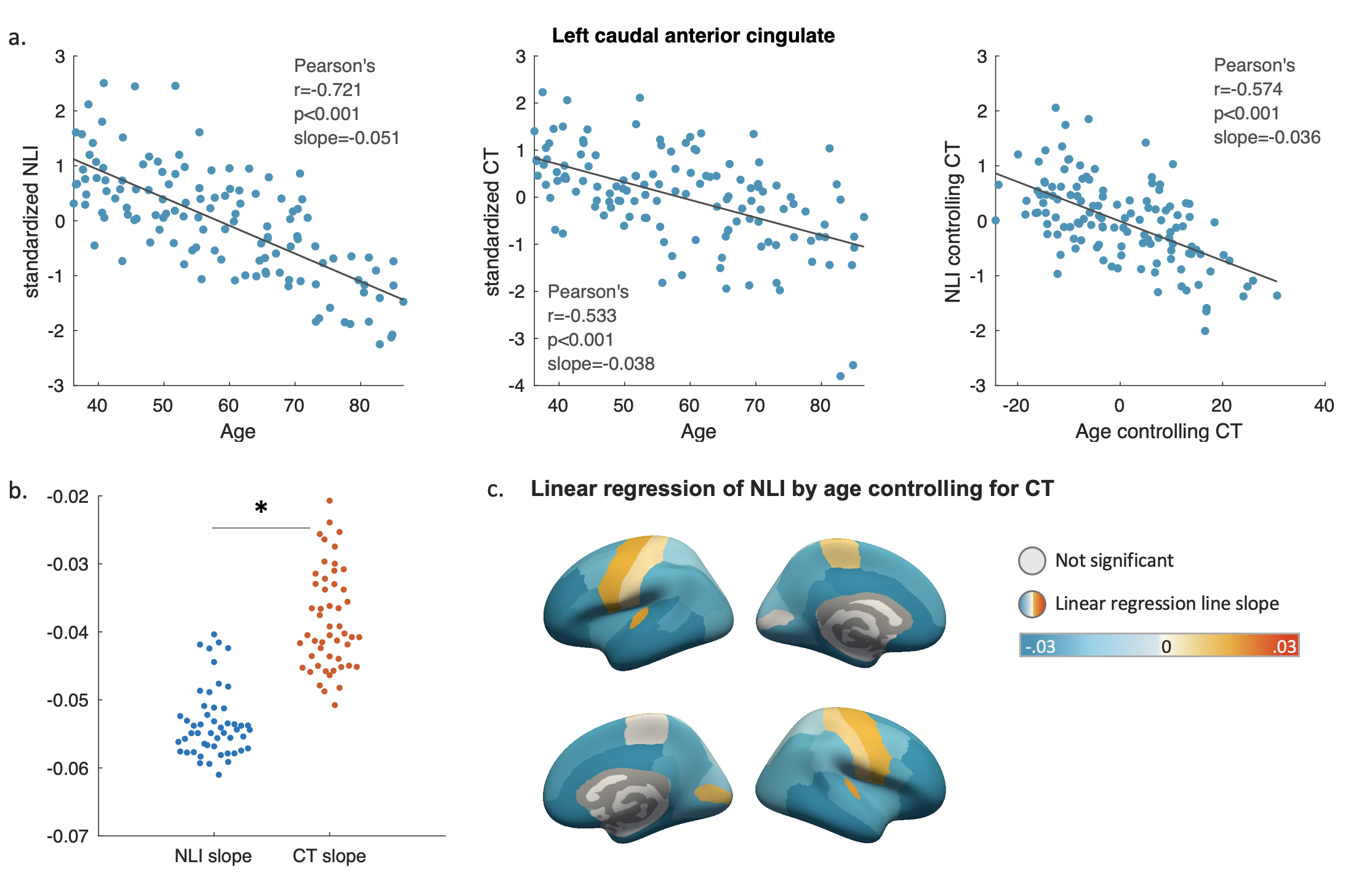
Figure 3. a) Scatter plots showing NLI and CT’s correlation with age in left caudal anterior cingulate. Partial plots of NLI against age controlling for CT are shown on the far right; b) The slopes of the linear fitting lines describing the decreases of standardized NLI and CT with age for all atlas regions. * indicates statistical significance; c) The slopes of linear fitting lines of the NLI as a function of age, controlling for CT. Color indicates slope values from significant partial correlations. Darker blue indicates faster NLI decline with age controlling for CT.
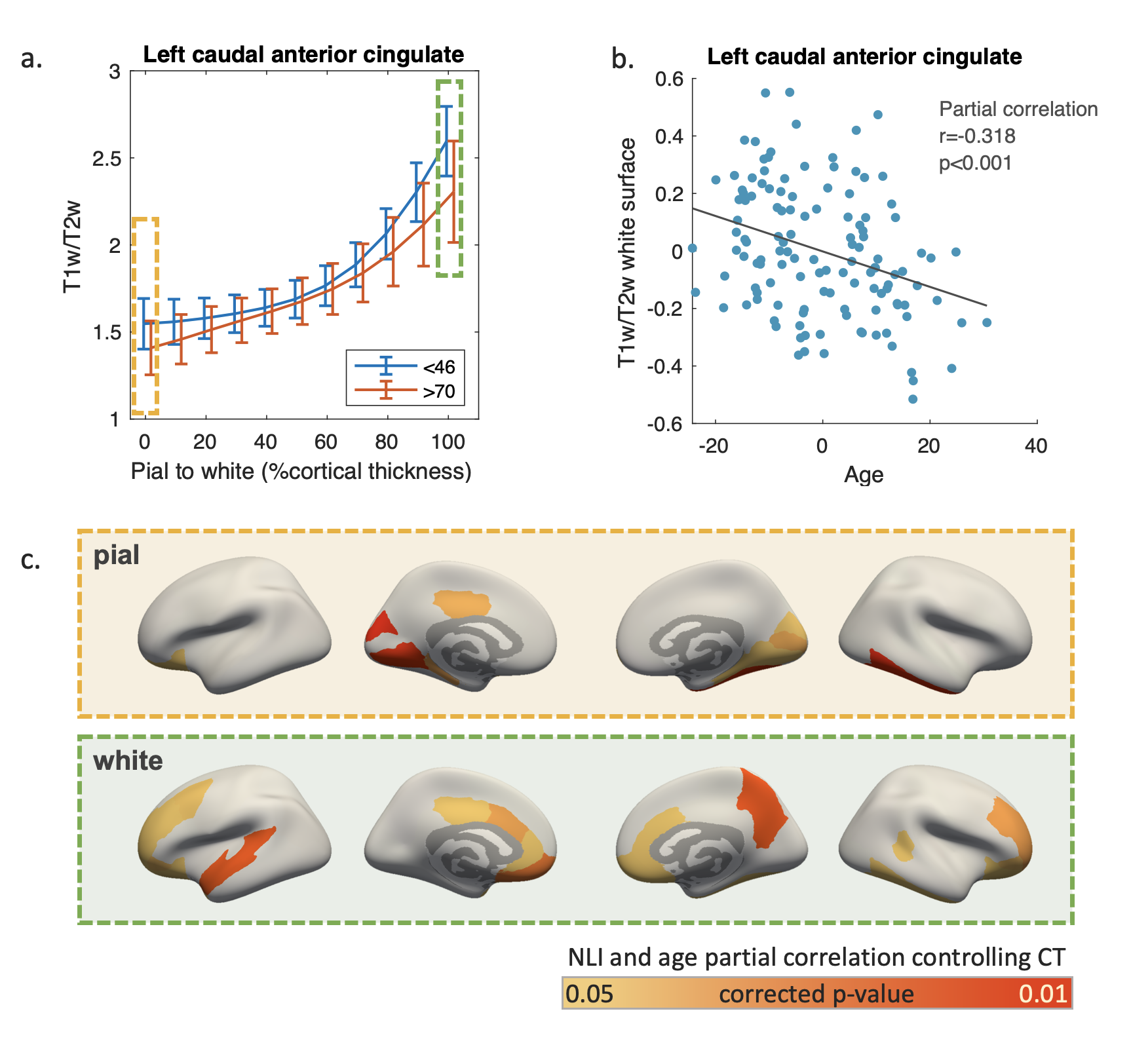
Figure 4. a) Sample T1w/T2w profiles of the youngest (blue) and oldest 25% participants (red) from the left caudal anterior cingulate region; b) Partial plot showing T1w/T2w value at white matter surface as a function of age after controlling for CT; c) Regions with significant age-related T1w/T2w decreases controlling for CT at pial and white matter surface. Color indicates the p-values of partial correlation after Bonferroni correction.