Grant S Roberts1, Leonardo A Rivera-Rivera2, Kevin M Johnson1,3, Sterling C Johnson2, Douglas C Dean III1,4, Andrew L Alexander1,5, Oliver Wieben1, and Laura B Eisenmenger3
1Medical Physics, University of Wisconsin - Madison, Madison, WI, United States, 2Medicine, University of Wisconsin - Madison, Madison, WI, United States, 3Radiology, University of Wisconsin - Madison, Madison, WI, United States, 4Pediatrics, University of Wisconsin - Madison, Madison, WI, United States, 5Psychiatry, University of Wisconsin - Madison, Madison, WI, United States
1Medical Physics, University of Wisconsin - Madison, Madison, WI, United States, 2Medicine, University of Wisconsin - Madison, Madison, WI, United States, 3Radiology, University of Wisconsin - Madison, Madison, WI, United States, 4Pediatrics, University of Wisconsin - Madison, Madison, WI, United States, 5Psychiatry, University of Wisconsin - Madison, Madison, WI, United States
NODDI and 4D flow MRI demonstrate significant
differences in white matter neurite density (NDI) in cognitively normal and
Alzheimer’s disease subjects, as well as correlations between cerebral blood
flow and NDI in cognitively normal subjects.
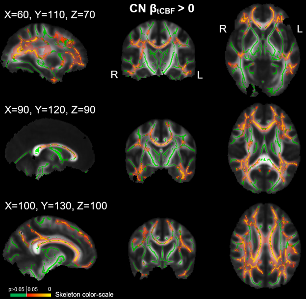
Tract-based spatial statistics (TBSS) analysis showing
regions on the white matter skeleton where the null hypothesis is rejected
(yellow with red filling; p≤0.05) and accepted (green; p>0.05). The null
hypothesis proposes that there exists no positive correlation between neurite
density (NDI) and total cerebral blood flow (tCBF) in cognitively normal (CN)
subjects. The mean fractional anisotropy image is used as the underlay.
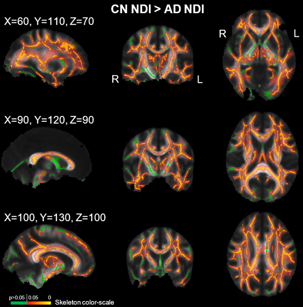
Tract-based spatial statistics (TBSS) analysis showing
regions on the white matter skeleton (at 3 MNI coordinates) where the null
hypothesis is rejected (yellow with red filling; p≤0.05) and accepted (green;
p>0.05). The null hypothesis proposes that neurite density (NDI) is not
greater in cognitively normal (CN) subjects compared to Alzheimer’s disease
(AD) subjects. The mean fractional anisotropy image is used as the underlay.
Note some areas were erroneously masked after TBSS registration.