Antoine Klauser1,2, Sebastien Courvoisier1,2, Michel Kocher1,2, and François Lazeyras1,2
1Radiology and Medical Informatics, University of Geneva, Geneva, Switzerland, 2CIBM Center for Biomedical Imaging, Geneva, Switzerland
1Radiology and Medical Informatics, University of Geneva, Geneva, Switzerland, 2CIBM Center for Biomedical Imaging, Geneva, Switzerland
Whole-brain High-resolution data were measured on volunteers with
a 1H 3D Adiabatic Spin-Echo MRSI sequence
and compared to FID-MRSI measurement.
The
overall
spectral quality are equivalent but usage of spin-echo enable further spectral editing technique.

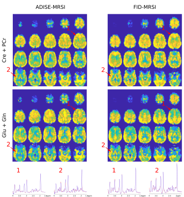
Comparison between 3D metabolite volumes resulting from ADISE-MRSI
and FID-MRSI acquisition sequences acquired subsequently in one
volunteer. Bottom, two sample spectra at same location for both
sequences are shown on the same scale and exhibit a slightly lower
signal with ADISE-MRSI sequence, consequence of the longer TE. Signal
loss due to B0 inhomogeneity in frontal lobe is present with both
sequences.

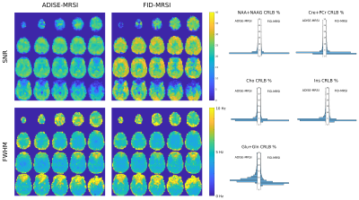
Comparison of spectral quality parameters between Fast ADISE-MRSI and
FID-MRSI sequences on the same session. Left, the signal loss due to
the longer TE for ADISE-MRSI is visible over the whole brain in the
SNR map. FWHM maps exhibits no marked difference. Right, the
histograms of the Cramer-Rao Lower Bound (CRLB) resulting from both sequences are displayed side to
side. Most metabolites show a slight shift towards higher CRLB values for
ADISE-MRSI. This is particularly visible for Glu+Gln that are known
to be more difficult to quantify at longer TE due to J-coupling
modulations.
