Manoj Kumar Sarma1,2, Andres Saucedo3, and M. Albert Albert Thomas3
1Advanced Imaging Research Center, UT Southwestern Medical Center, Dallas, TX, United States, 2Radiology, UT Southwestern Medical Center, Dallas, TX, United States, 3Radiology, UCLA School of Medicine, Los Angeles, CA, United States
1Advanced Imaging Research Center, UT Southwestern Medical Center, Dallas, TX, United States, 2Radiology, UT Southwestern Medical Center, Dallas, TX, United States, 3Radiology, UCLA School of Medicine, Los Angeles, CA, United States
We propose an echo planar-based
diffusion weighted spectroscopic imaging using semi‐LASER localization and
bipolar diffusion gradient. Initial results show good spectral quality and spatial
localization.
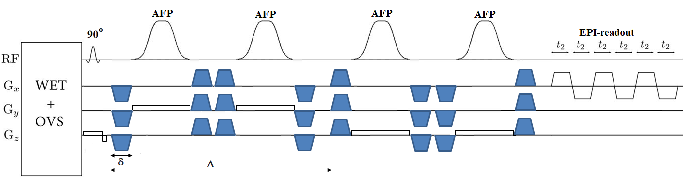
Figure 1: Schematic
representation of the DW-sL-EPSI sequence showing RF pulses with bipolar DW
gradients placed around the slice selective refocusing HS RF pulses. δ = gradient duration, sum of the durations of four lobes; Δ = diffusion time, time between the first lobe of the
dephasing diffusion gradient group and the first lobe of the re-phasing
diffusion gradient group. Water suppression was performed with WET. An EPI-based
readout was used to capture the k-t data.
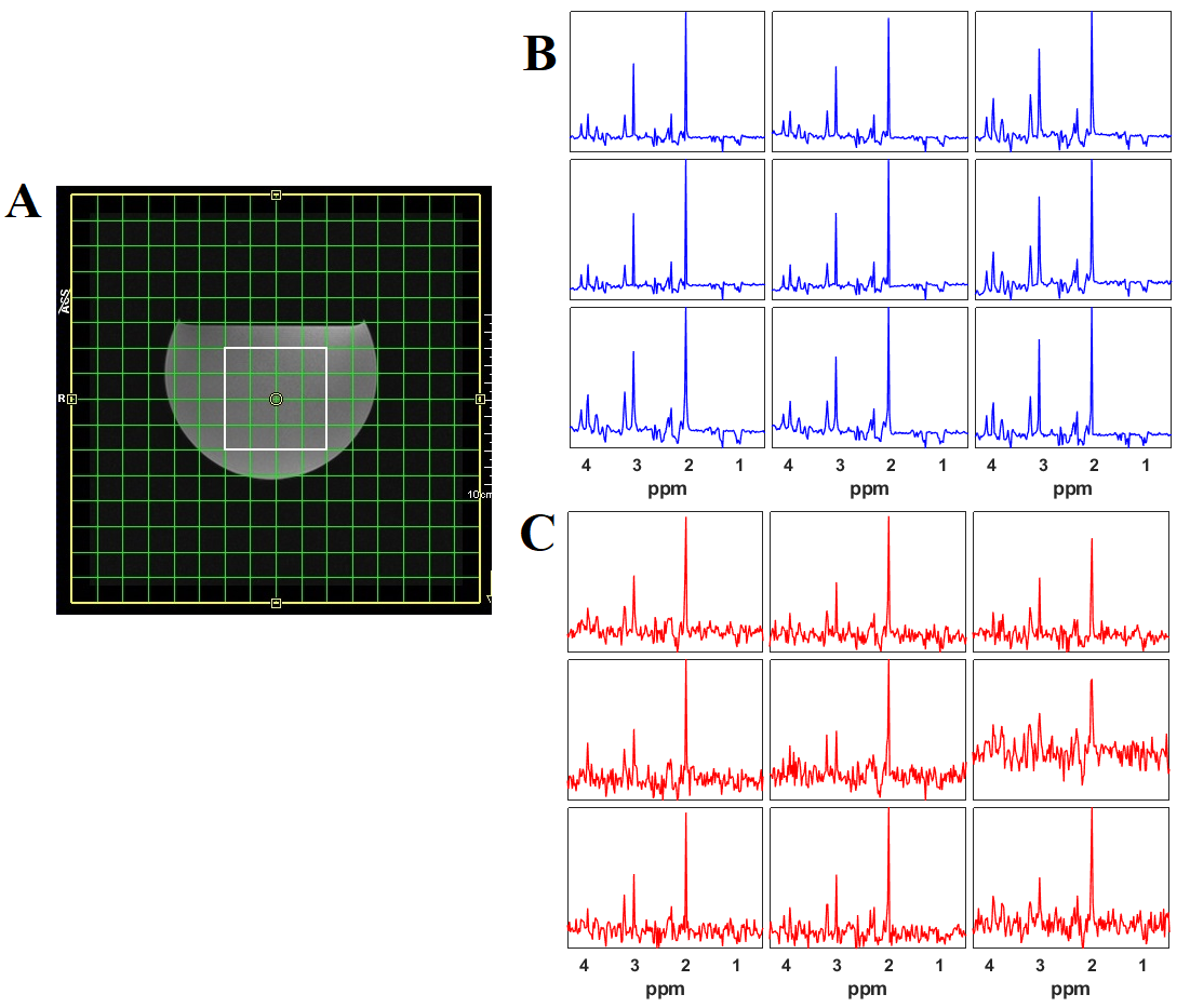
Figure 2: Results from a brain phantom scan. (A) T1-weighted localization
image showing the VOI; also shown is the DW-sL-EPSI spectra obtained from the 3x3
region within the VOI at the b-values (B) 36 s/mm2 and (C)
3996 s/mm2.