Odélia Jacqueline Chitrit1, Qingjia Bao1, Maxime Yon1, and Lucio Frydman1
1Department of Chemical and Biological Physics, Weizmann institute of Science, Rehovot, Israel
1Department of Chemical and Biological Physics, Weizmann institute of Science, Rehovot, Israel
The present study explores the use of a customized 3D phase-encoded Spatiotemporal Encoding (SPEN) MRI approach delivering quality DTI volumetric data at 15.2T of mice fetal brains in utero, as well as within the first week post-partum.
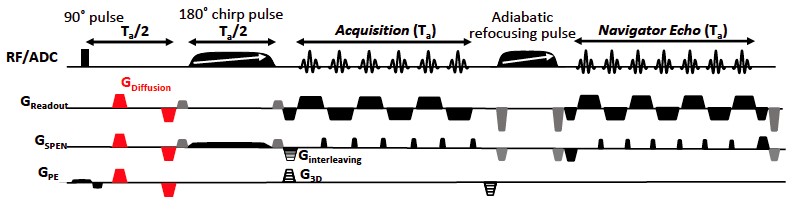
Figure 1: Sequence employed for the 3D
SPEN DTI acquisitions, including a 180˚ chirp pulse acting in the presence
of a gradient that encodes the more artifact-prone, low bandwidth dimension, a
pre-encoding Ta/2 delay introduced for achieving SPEN’s
full-refocusing condition where variable-orientation diffusion-weighting
gradients (red) are placed, and gradients for interleaving and phase-encoding
procedures. A final, 2D echo-based reacquisition of the k=0 PE line was
included for all interleaves, for correcting motion phase distortions in these
experiments.
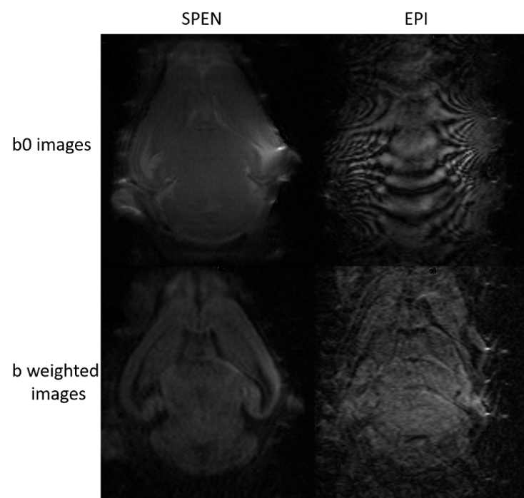
Figure 3: Comparison between
in vivo EPI and SPEN images extracted from 3D DTI acquisitions on a 2-days-old live
mouse. The in-plane fields targeted in both experiments were 10x10 mm.