Chenyang Li1,2, Els Fieremans1, Dmitry S. Novikov1, Yulin Ge1, and Jiangyang Zhang1
1Department of Radiology, Center for Biomedical Imaging, NYU Grossman School of Medicine, New York, NY, United States, 2Vilcek Institute of Graduate Biomedical Sciences, NYU Grossman School of Medicine, New York, NY, United States
1Department of Radiology, Center for Biomedical Imaging, NYU Grossman School of Medicine, New York, NY, United States, 2Vilcek Institute of Graduate Biomedical Sciences, NYU Grossman School of Medicine, New York, NY, United States
In this study, water exchange effects in postmortem mouse brains were measured using Filter Exchange Imaging (FEXI) and diffusion time dependent kurtosis imaging (td-DKI) and a correlation has been observed between them, suggesting that they are sensitive to similar exchange processes.
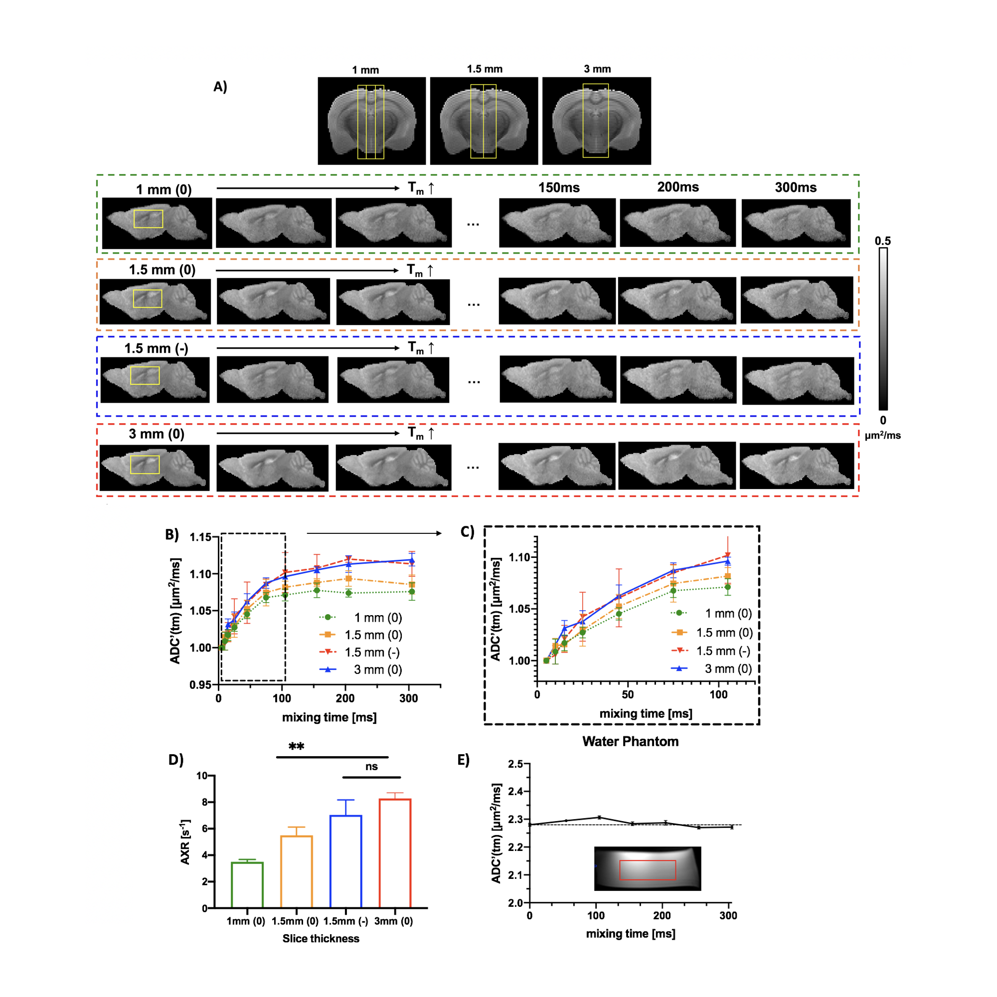
Figure 1. A) Representative ADC’(tm) map in slice thickness of 1mm, 1.5mm and 3mm with no crusher and 1.5mm with negative crusher. B) Normalized ADC’(tm)-vs-tm from 5 ms to 305 ms in ROI of brain (yellow). C) Normalized ADC’(tm)-vs-tm with mixing time from 5 ms to 105 ms. D) AXR estimated from 5-105 ms. E) ADC’(tm) in a phantom with filter gradient and readout gradient as 100 s/mm2.
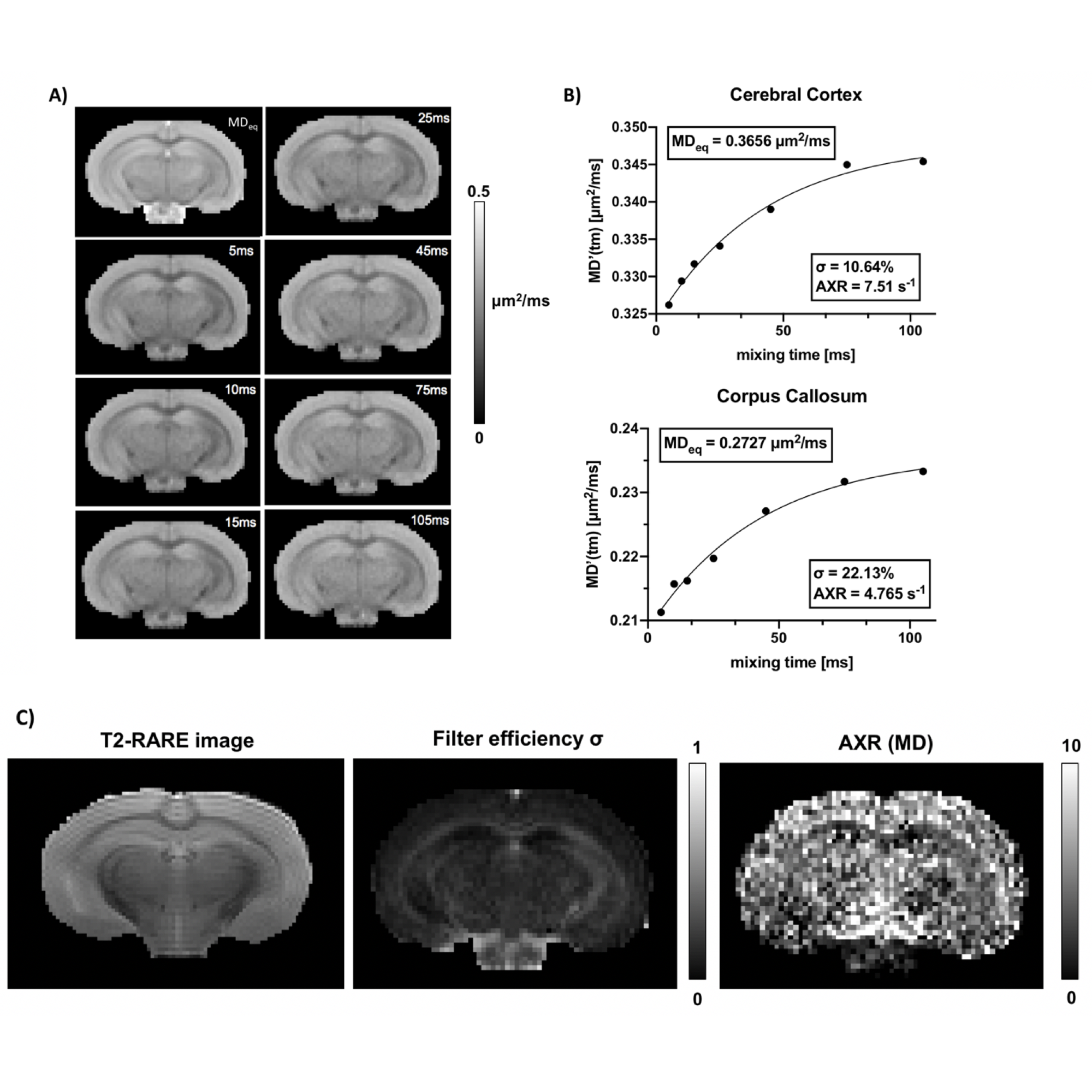
Figure 2. A) Representative MD’(tm) map with mixing time from 5 ms to 105 ms. B) MD’(tm)-vs-tm curve in cerebral cortex and corpus callosum with mixing time from 5 ms to 105 ms. C) shows the structural reference T2-RARE images along with fitted voxel-wise filter efficiency map and AXR with mixing time from 5 ms to 105 ms.