Mi Zhou1, Robert Stobbe1, Filip Szczepankiewicz2,3, Mar Lloret4, Brian Buck4, Paige Fairall4, Ken Butcher4, Ashfaq Shuaib4, Derek Emery5, Markus Nilsson2, Carl-Fredrik Westin3, and Christian Beaulieu1
1Biomedical Engineering, University of Alberta, Edmonton, AB, Canada, 2Clinical Sciences Lund, Lund University, Lund, Sweden, 3Radiology, Brigham and Women’s Hospital, Harvard Medical School, Boston, MA, United States, 4Neurology, University of Alberta, Edmonton, AB, Canada, 5Radiology and Diagnostic Imaging, University of Alberta, Edmonton, AB, Canada
1Biomedical Engineering, University of Alberta, Edmonton, AB, Canada, 2Clinical Sciences Lund, Lund University, Lund, Sweden, 3Radiology, Brigham and Women’s Hospital, Harvard Medical School, Boston, MA, United States, 4Neurology, University of Alberta, Edmonton, AB, Canada, 5Radiology and Diagnostic Imaging, University of Alberta, Edmonton, AB, Canada
A rapid multidimensional
b-tensor encoding protocol was applied to acute stroke patients showing higher
microscopic anisotropy and tissue heterogeneity in ischemic white and grey
matter, but with a concomitant decrease in typical DTI fractional anisotropy.
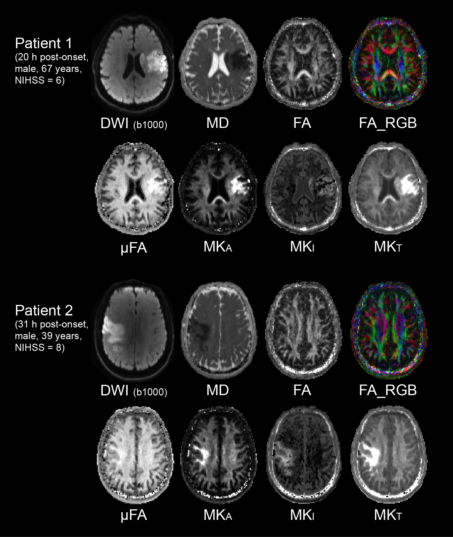
Figure
2: Raw DWI b1000 (pre-scan normalize off) and smoothed
b-tensor-derived diffusion metric maps are shown for two acute stroke patients.
In both cases, the low region of mean diffusivity (MD) shows no change on
standard tensor fractional anisotropy (FA), but greatly elevated microscopic FA
(µFA), anisotropic, isotropic, and total kurtosis (MKA, MKI, MKT)
within the ischemic white matter.
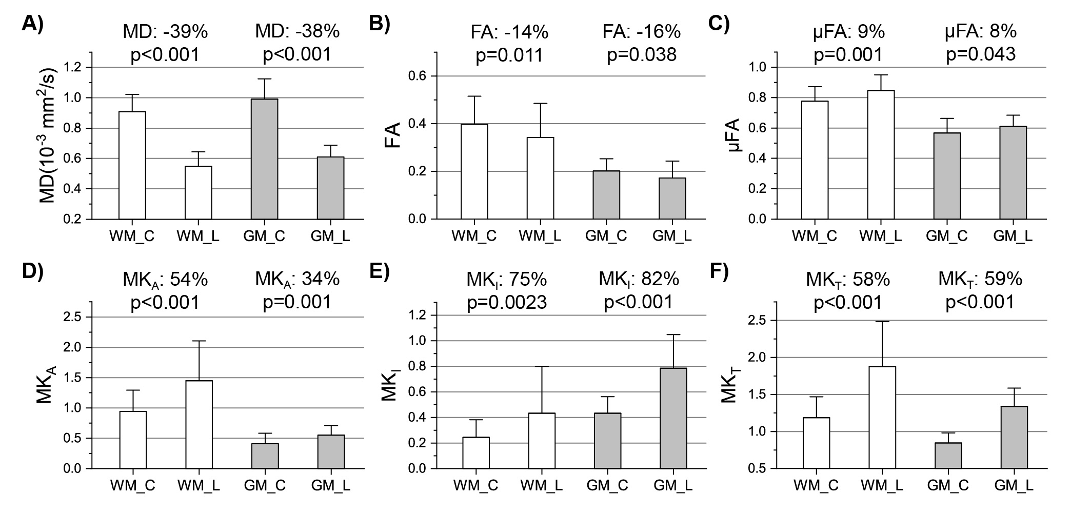
Figure
3: The mean ± SD over 21 acute stroke patients are
shown for regions over multiple slices of WM and GM within the lesion (‘L’) and
contralateral (‘C’) brain. (A) Lesion MD was reduced by 39% in WM and 38% in GM.
(B) FA was 14% lower in the lesion relative to contralateral WM. (C-F) In
contrast, the b-tensor derived parameters were all elevated in the lesion WM by
9% for microscopic FA (µFA), 54%
for anisotropic kurtosis (MKA), 75% for isotropic kurtosis
(MKI), and 58% for total kurtosis (MKT). Similar changes
were found in ischemic GM.