Gabriel Ramos-Llordén1, Chiara Maffei1, Qiyuan Tian1, Berkin Bilgic1,2, Thomas Witzel3, Boris Keil4, Anatasia Yendiki1, and Susie Huang1,2
1Athinoula A. Martinos Center for Biomedical Imaging, Department of Radiology, Masachusetts General Hospital, Harvard Medical School, Charlestown, MA, United States, 2Harvard-MIT Division of Health Sciences and Technology, Massachusetts Institute of Technology, Cambridge, MA, United States, 3Q Bio Inc, San Carlos, CA, United States, 4Institute of Medical Physics and Radiation Protection, Mittelhessen University of Applied Sciences, Giessen, Germany
1Athinoula A. Martinos Center for Biomedical Imaging, Department of Radiology, Masachusetts General Hospital, Harvard Medical School, Charlestown, MA, United States, 2Harvard-MIT Division of Health Sciences and Technology, Massachusetts Institute of Technology, Cambridge, MA, United States, 3Q Bio Inc, San Carlos, CA, United States, 4Institute of Medical Physics and Radiation Protection, Mittelhessen University of Applied Sciences, Giessen, Germany
High b value diffusion MRI (up to 10 000 s / mm2) at ultra-high spatial resolution (550 micrometer) reveals highly detailed anatomical and connectivity information in relevant structure of an ex-vivo whole human brain.
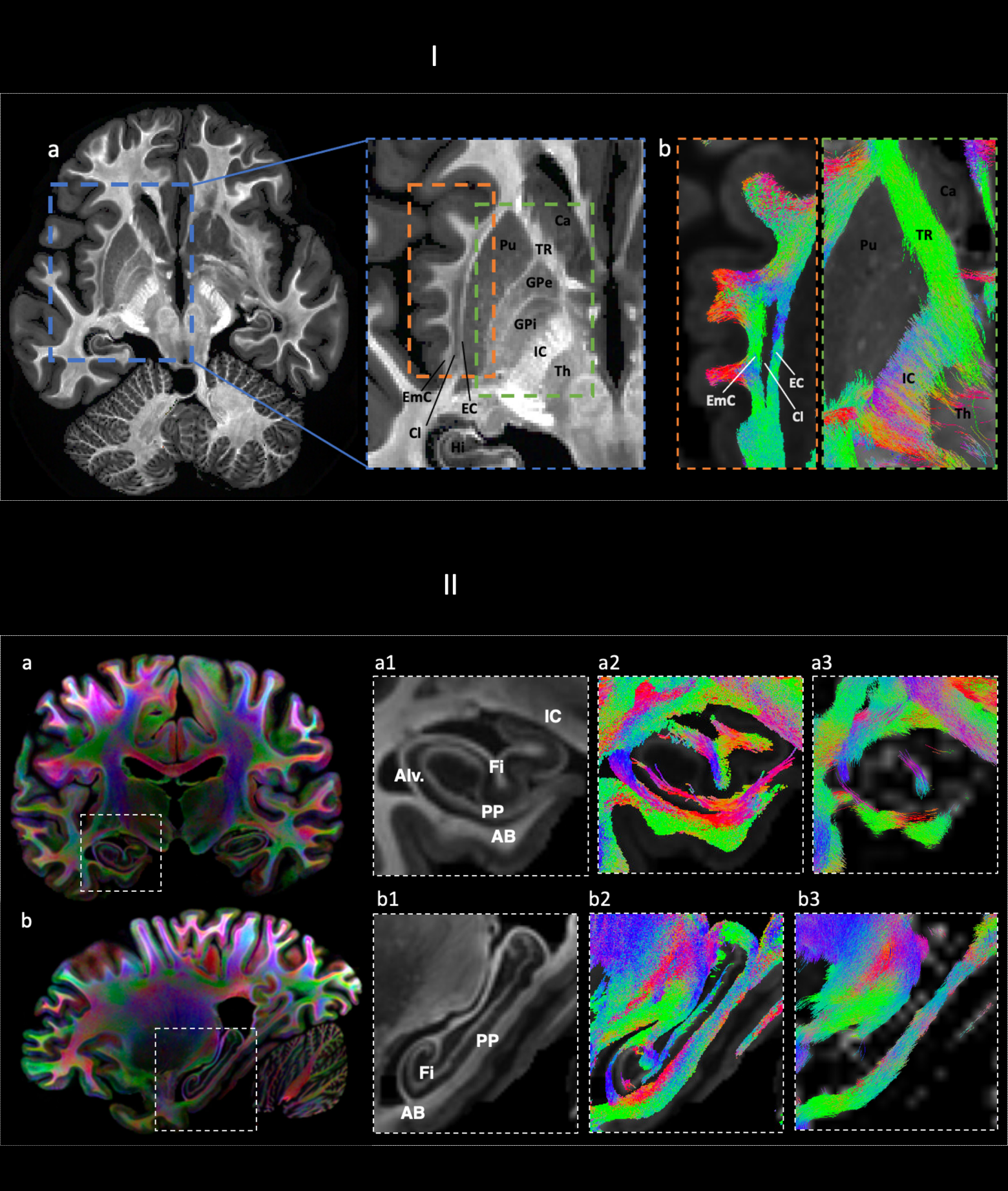
I. a) Mean kurtosis b) tractography showing branching between EC and EmC (left) and thalamic connectivity (right). Ca: caudate. Cl: claustrum. EC: external capsule. Emc: extreme capsule. IC: internal capsule. GPi/GPe: internal/external globus pallidus. Th: thalamus. Pu: putamen. TR: thalamic radiation.
II a-b) CSD a-b1,2,3: in a-b. 1) CSD-based maps (0-th order SH) showing main hippocampal formation structures. 2) tractography showing internal hippocampal connectivity. 3) lower spatial resolution .
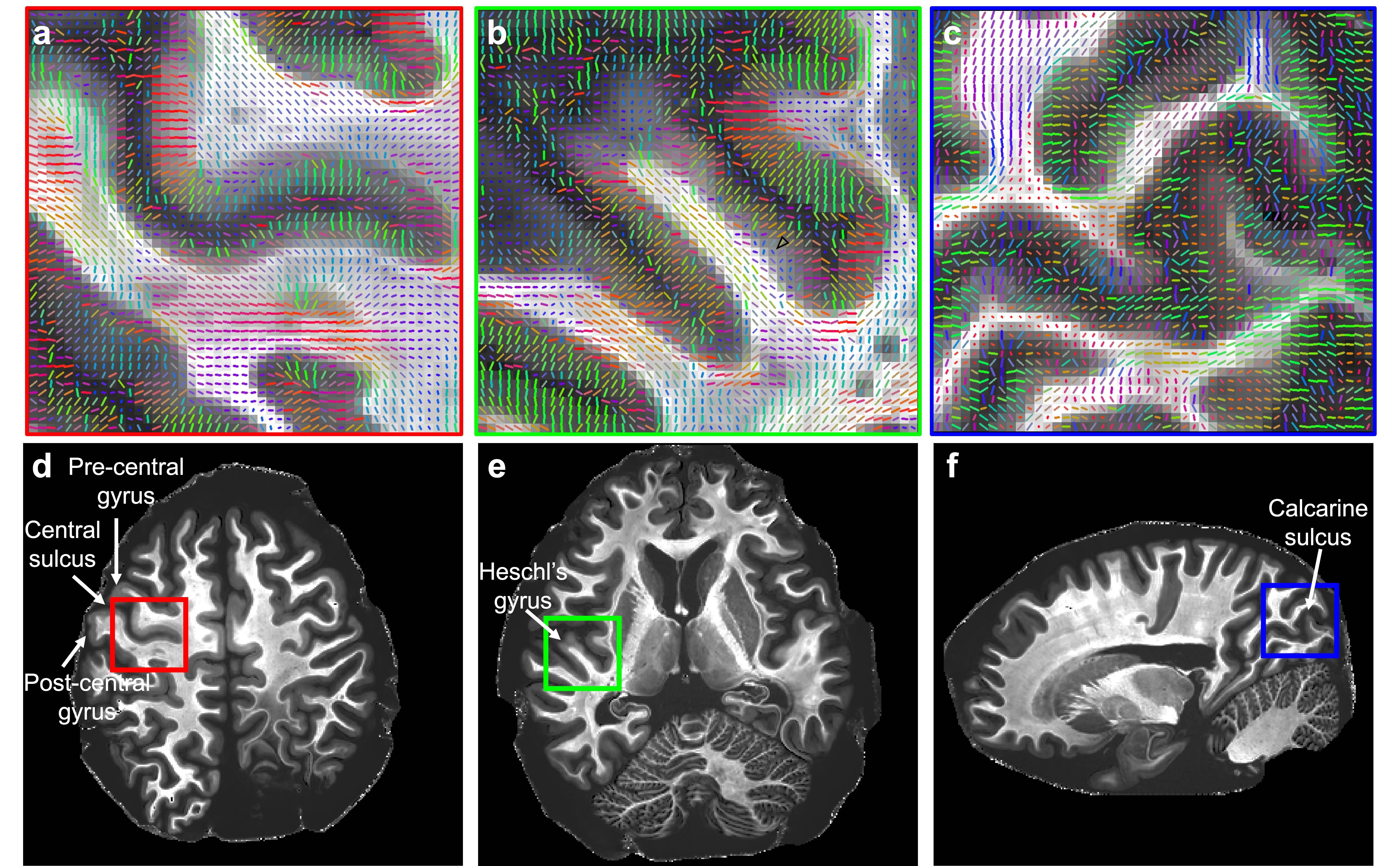
Primary eigenvectors of diffusion tensor imaging on top of mean kurtosis maps (a-c) from granular cortices where cortical fibers that are tangential to the cortical surface exist, shown in regions of interest selected from the primary somatosensory (d, red box), primary auditory (e, green box) and primary visual (c, blue box) cortex.