Yue Wu1,2,3, Chengyue Sun4, Qingle Kong5, Zhixin Li1,2,3, Dongbiao Sun1,2,3, Chen Ling4, Jing An6, Rong Xue1,2,3, Yan Zhuo1,2,3, Yun Yuan4, and Zihao Zhang1,2,3
1State Key Laboratory of Brain and Cognitive Science, Institute of Biophysics, Chinese Academy of Sciences, Beijing, China, 2The Innovation Center of Excellence on Brain Science, Chinese Academy of Sciences, Beijing, China, 3University of Chinese Academy of Sciences, Beijing, China, 4Department of Neurology, Peking University First Hospital, Beijing, China, 5MR Collaboration, Siemens Healthcare Ltd, Beijing, China, 6Siemens Shenzhen Magnetic Resonance Ltd, Shenzhen, China
1State Key Laboratory of Brain and Cognitive Science, Institute of Biophysics, Chinese Academy of Sciences, Beijing, China, 2The Innovation Center of Excellence on Brain Science, Chinese Academy of Sciences, Beijing, China, 3University of Chinese Academy of Sciences, Beijing, China, 4Department of Neurology, Peking University First Hospital, Beijing, China, 5MR Collaboration, Siemens Healthcare Ltd, Beijing, China, 6Siemens Shenzhen Magnetic Resonance Ltd, Shenzhen, China
Phase-contrast MR angiography at 7T was proved to be a reliable,
non-invasive method to evaluate small arterial dysfunction in cerebral small vessel
disease (CSVD). Lenticulostriate arteries had decreased blood flow velocities, and
were associated with MRI lesions and clinical symptoms.
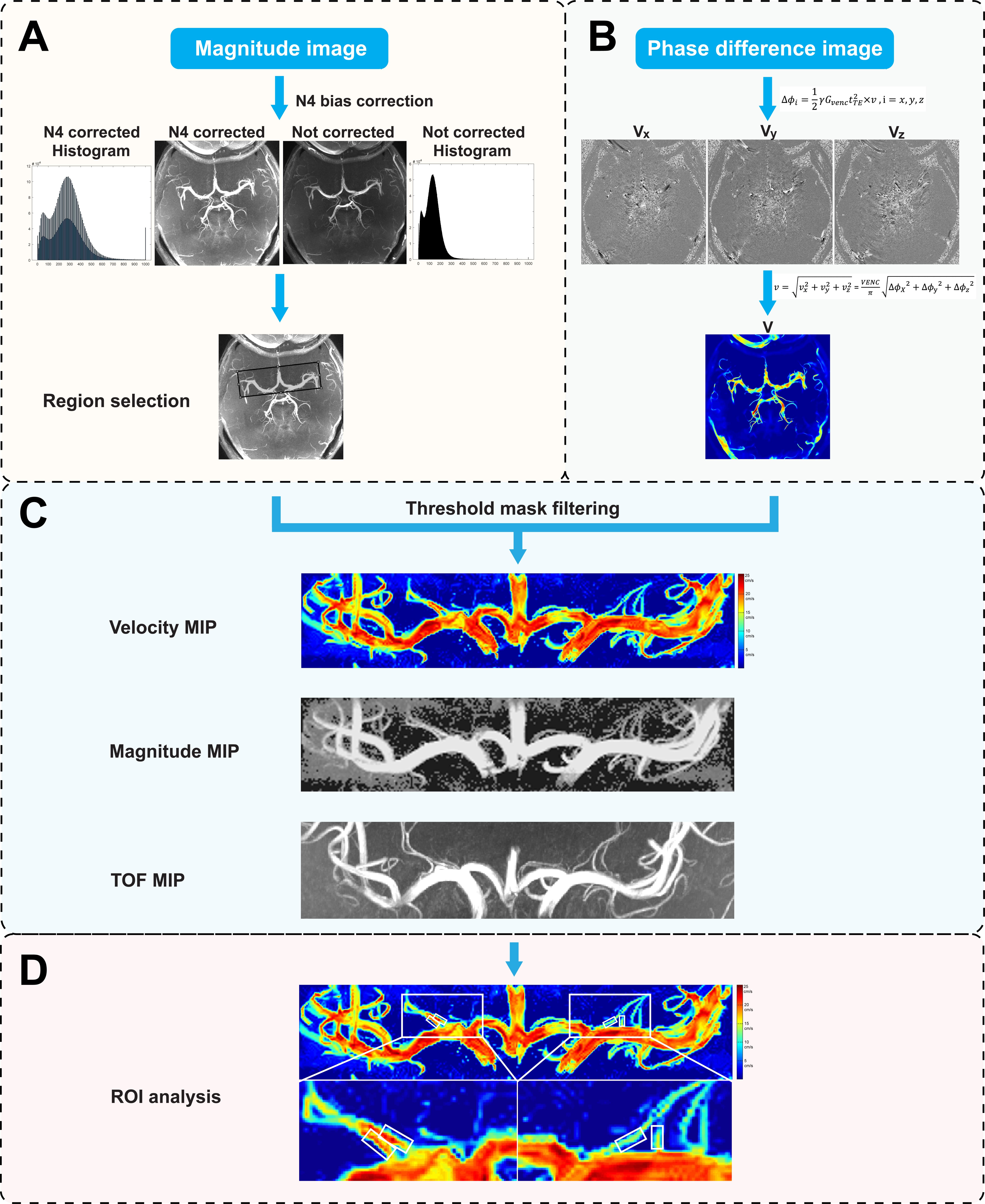
Figure
1. Data analysis workflow to quantify lenticulostriate
arterial (LSA) blood flow velocities (BFVs). (A) The reference
scan magnitude images after an N4 bias correction were threshold-filtered to
generate vascular mask. (B) The velocity dataset was calculated from phase-difference
images in three directions (Vx, Vy, Vz). (C) A mask was applied, and the
velocity map was reconstructed with maximal intensity projections (MIPs). (D)
Rectangular regions of interests (ROIs) with fixed sizes were placed on the MIP
images to measure LSA BFVs.
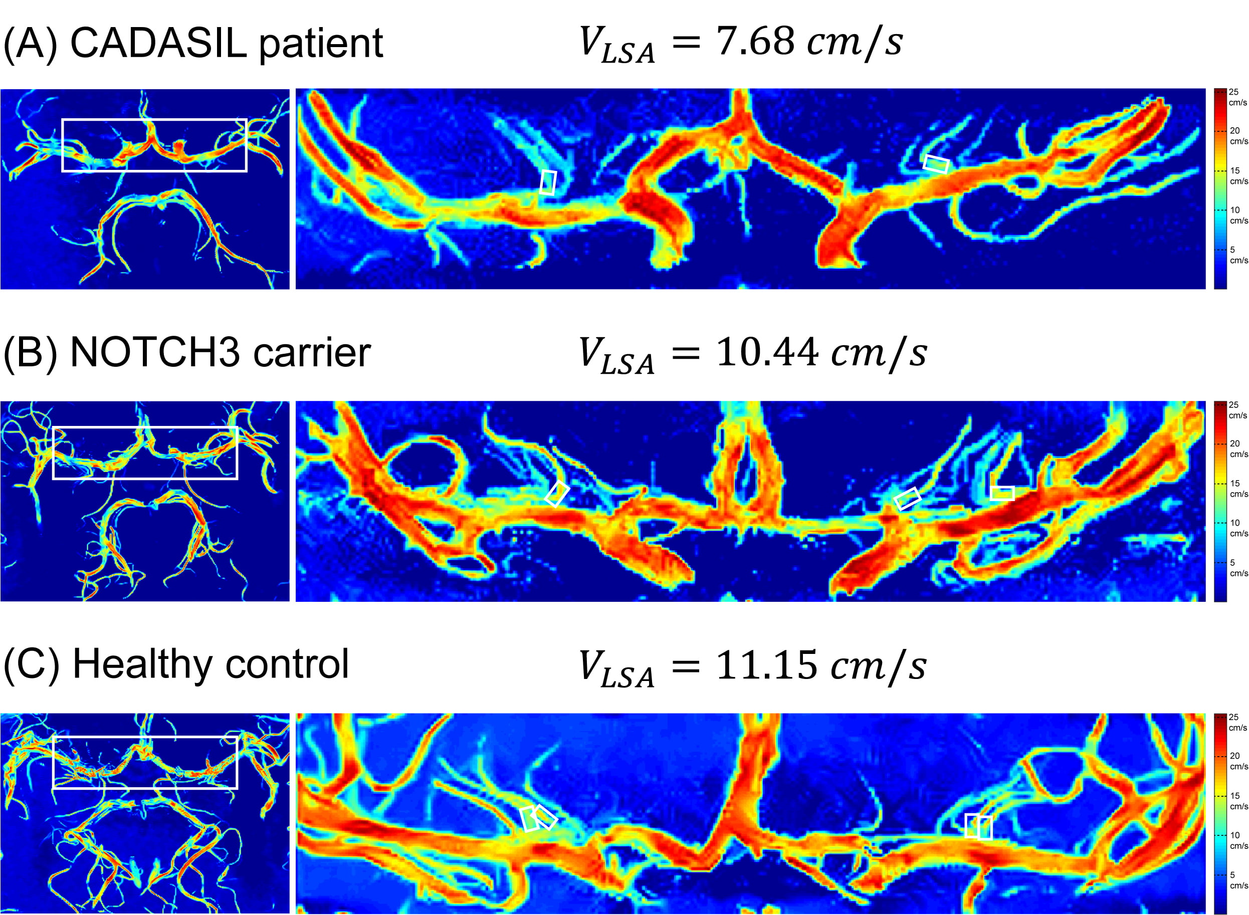
Figure
2. A representative case of symptomatic patient with cerebral
autosomal dominant arteriopathy with subcortical infarcts and leukoencephalopathy
(CADASIL) showing
reduced lenticulostriate arterial
(LSA) blood flow velocities. Velocity map of (A) a CADASIL patient;
(B) an age- and sex-matched asymptomatic NOTCH3 mutation carrier; and (C) an
age- and sex-matched healthy control.