Esteban Jorge Denecken-Campaña1,2,3, Julio Sotelo1,3,4, Cristobal Arrieta1,3, Pablo Irarrazaval1,2,3, Cristián Tejos1,2,3, Marcelo E. Andia1,3,5, and Sergio Uribe1,3,5
1Biomedical Imaging Center, Pontificia Universidad Católica de Chile, Santiago, Chile, 2Electrical Engineering Department, School of Engineering, Pontificia Universidad Católica de Chile, Santiago, Chile, 3ANID – Millennium Science Initiative Program – Millennium Nucleus for Cardiovascular Magnetic Resonance, Santiago, Chile, 4School of Biomedical Engineering, Universidad de Valparaíso, Valparaíso, Chile, 5Department of Radiology, School of Medicine, Pontificia Universidad Católica de Chile, Santiago, Chile
1Biomedical Imaging Center, Pontificia Universidad Católica de Chile, Santiago, Chile, 2Electrical Engineering Department, School of Engineering, Pontificia Universidad Católica de Chile, Santiago, Chile, 3ANID – Millennium Science Initiative Program – Millennium Nucleus for Cardiovascular Magnetic Resonance, Santiago, Chile, 4School of Biomedical Engineering, Universidad de Valparaíso, Valparaíso, Chile, 5Department of Radiology, School of Medicine, Pontificia Universidad Católica de Chile, Santiago, Chile
Hemodynamic parameters from 4D flow datasets acquired with self-gating acquisition showed statistically significant differences compared to those measured without self-gating. We found significant variability of these parameters in the ascending aorta when comparing both methods.
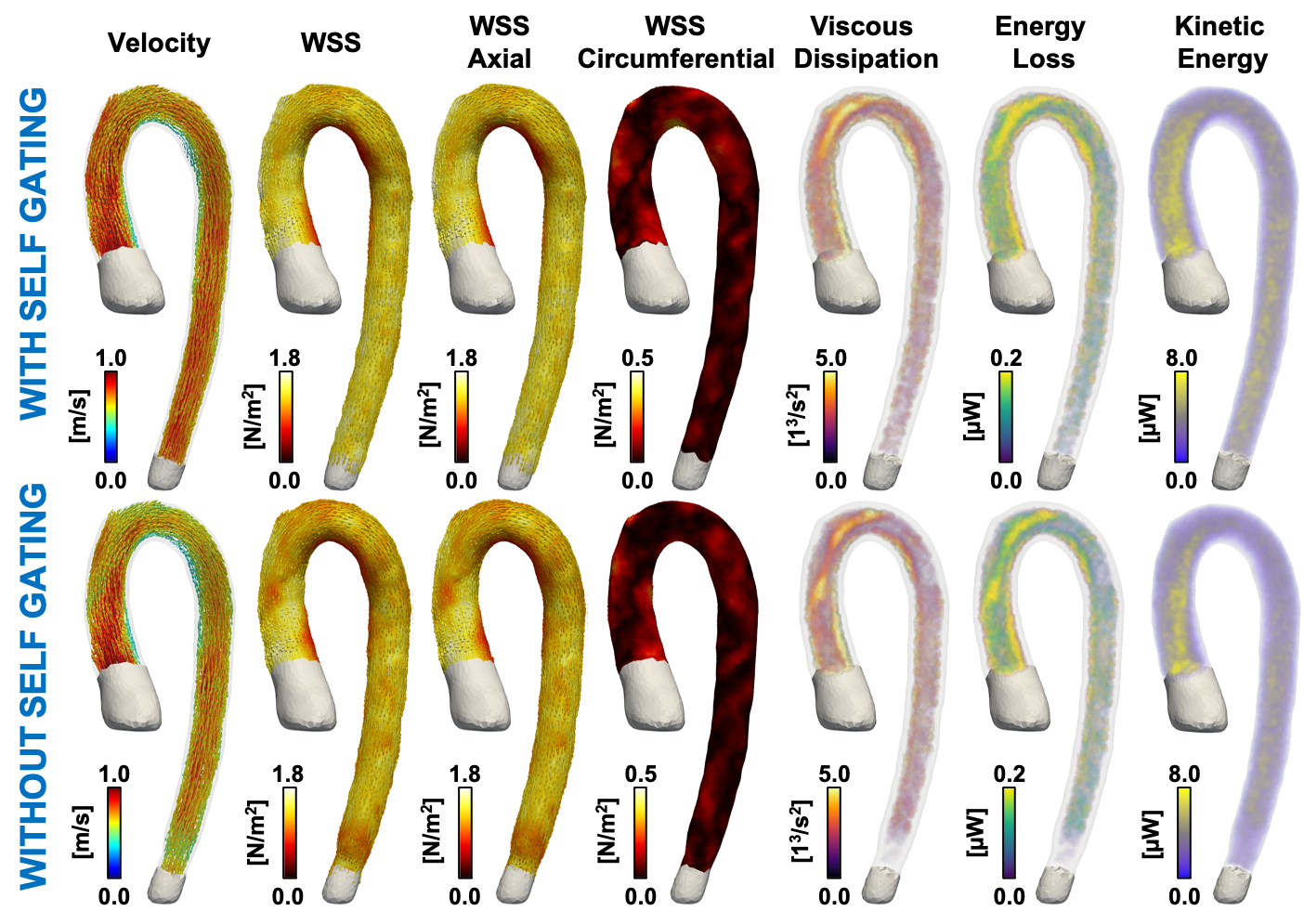
Hemodynamic parameters obtained from 4D flow MRI with and without self-gating. In this figure we show visual representations of the hemodynamic parameters for one volunteer.
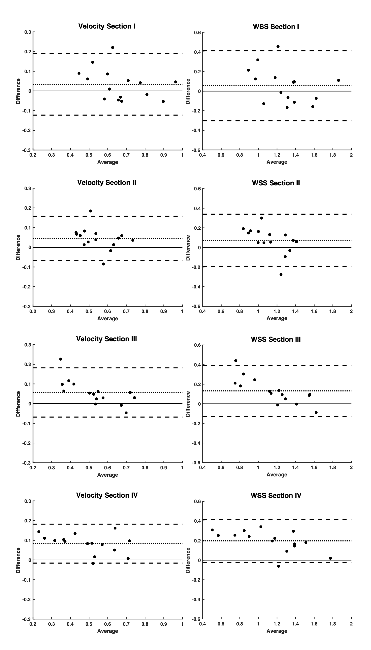
Bland Altman plots of velocity and wall shear stress at each section. The difference in Bland Altman plots is computed as: SG—NG, where SG is the data with self-gating and NG is the data without self-gating.