Jian Hou1, Vincent Wai-Sun Wong2, Grace Lai-Hung Wong2, Baiyan Jiang1, Yi-Xiang Wang1, Anthony Wing-Hung Chan3, Winnie Chiu-Wing Chu1, and Weitian Chen1
1Department of Imaging and Interventional Radiology, The Chinese University of Hong Kong, Hong Kong, Hong Kong, 2Department of Medicine & Therapeutics, The Chinese University of Hong Kong, Hong Kong, Hong Kong, 3Department of Anatomical and Cellular Pathology, The Chinese University of Hong Kong, Hong Kong, Hong Kong
1Department of Imaging and Interventional Radiology, The Chinese University of Hong Kong, Hong Kong, Hong Kong, 2Department of Medicine & Therapeutics, The Chinese University of Hong Kong, Hong Kong, Hong Kong, 3Department of Anatomical and Cellular Pathology, The Chinese University of Hong Kong, Hong Kong, Hong Kong
MPF measured using MPF-SL shows correlations with
the degree of liver fibrosis. A significant difference of MPF between normal
(F0) and early stage liver fibrosis (F1/F2) was observed.
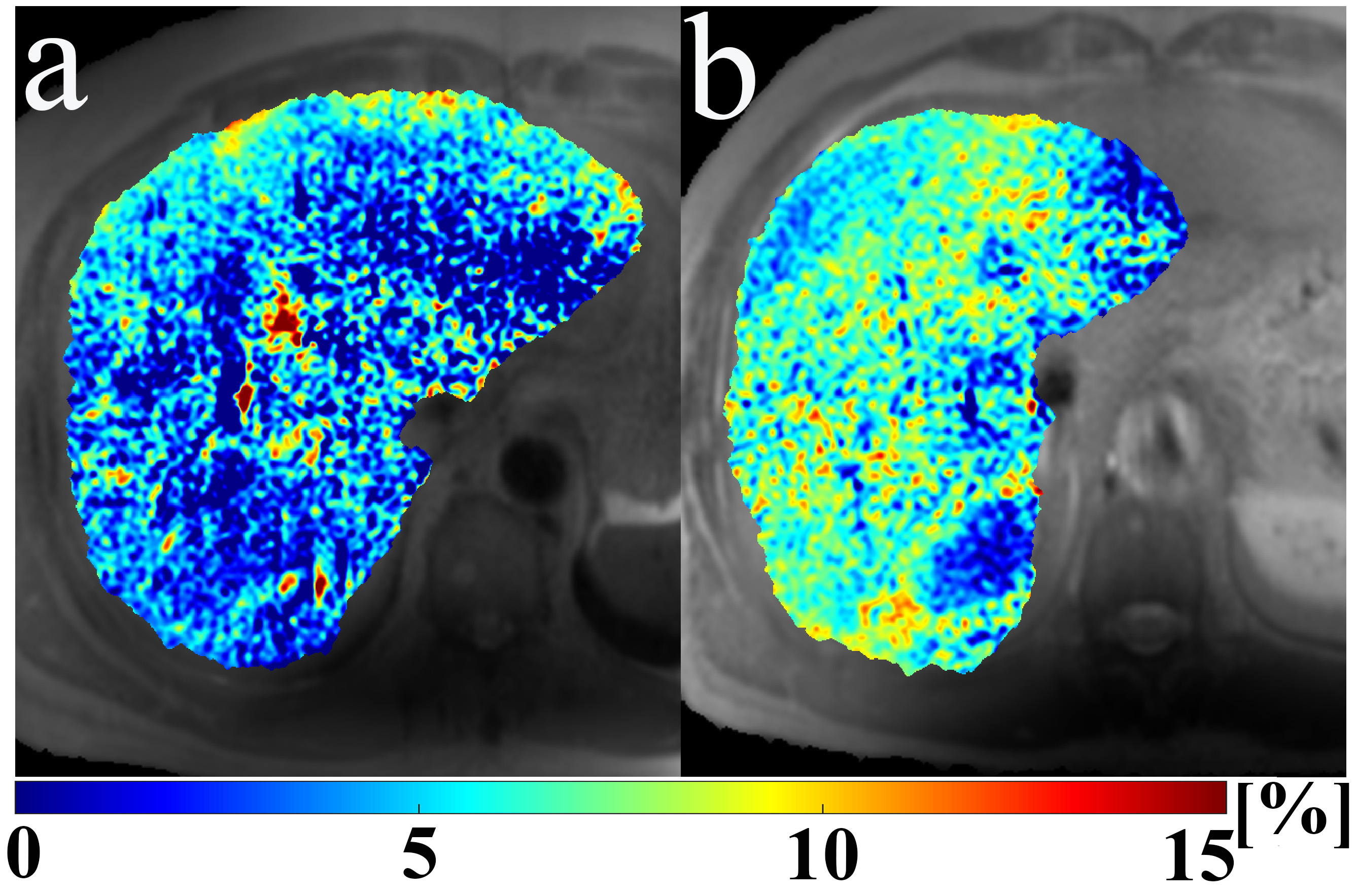
Figure 1. Example MPF
maps, a): from a patient without liver fibrosis (F0), and b): from a patient
with fibrosis stage F2. All results were confirmed by liver biopsy.
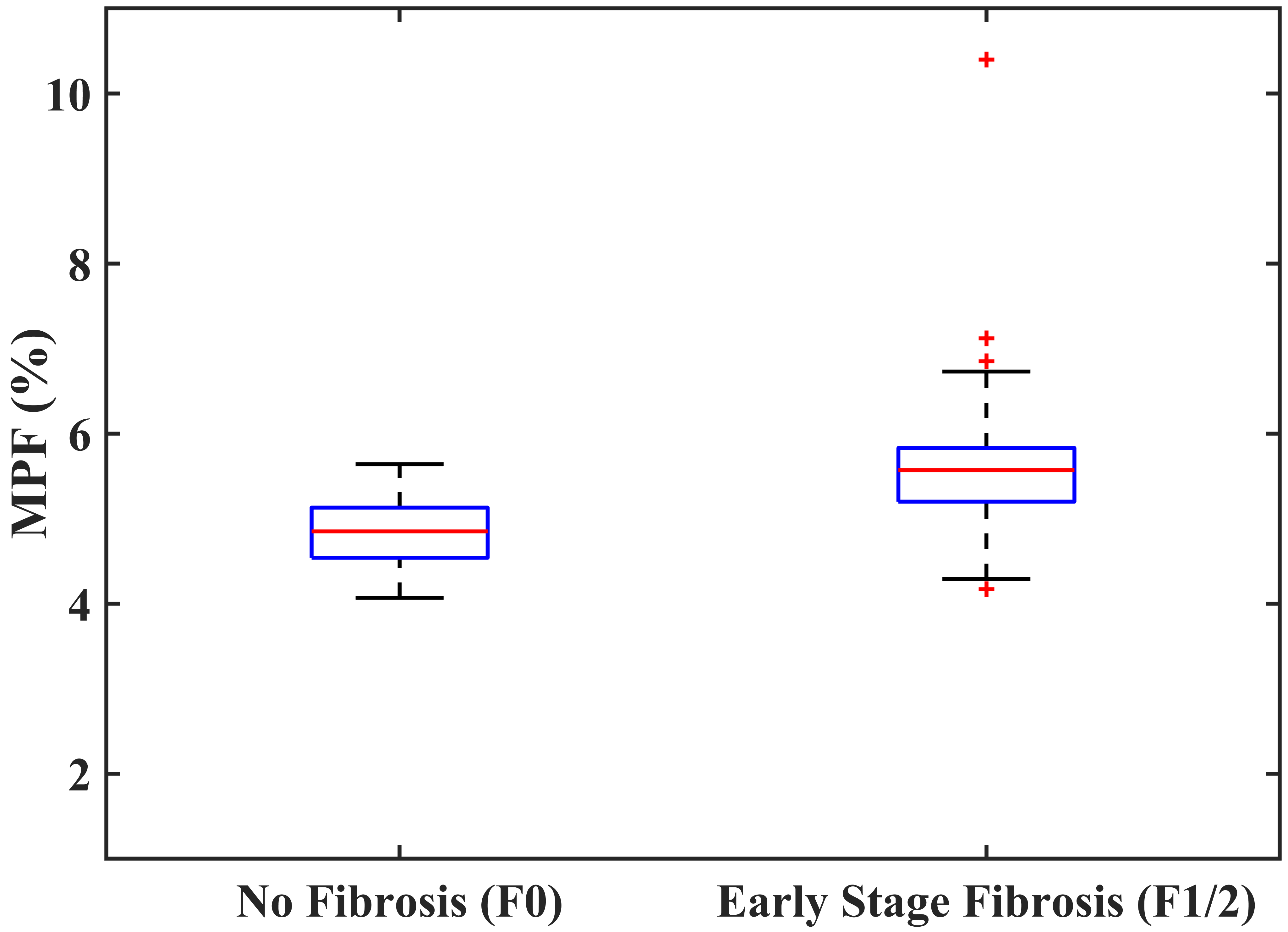
Figure 2. The box plot
of the measured MPF using MPF-SL at stage F0 and stage F1/F2.