Fasil Gadjimuradov1,2, Thomas Benkert2, Marcel Dominik Nickel2, and Andreas Maier1
1Pattern Recognition Lab, Friedrich-Alexander-Universität Erlangen-Nürnberg, Erlangen, Germany, 2Magnetic Resonance, Siemens Healthcare GmbH, Erlangen, Germany
1Pattern Recognition Lab, Friedrich-Alexander-Universität Erlangen-Nürnberg, Erlangen, Germany, 2Magnetic Resonance, Siemens Healthcare GmbH, Erlangen, Germany
Partial Fourier acquisitions can be used to increase SNR in DWI. While conventional techniques introduce artifacts in liver DWI due to smoothness priors of the phase, this work proposes to reconstruct using a recurrent neural network implementing a more suitable regularization.
Figure
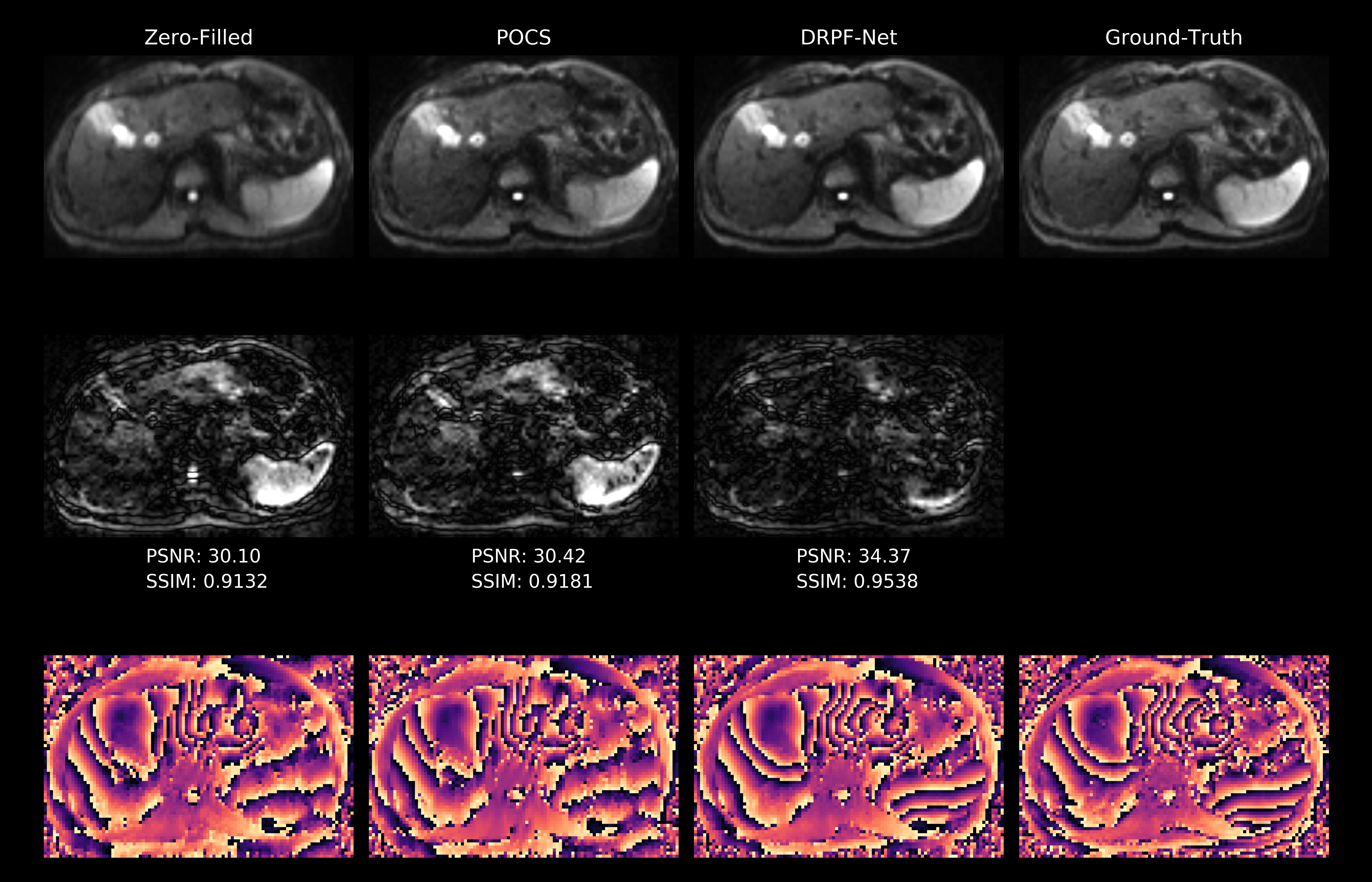
3: Qualitative comparison of zero-filling, POCS and DRPF-Net for a combined DW
liver image (b = 800 s/mm2). First row: the zero-filled
reconstruction is blurred along AP, while POCS exhibits artificial,
ringing-like structures most apparent in the left liver lobe. In contrast, the
DRPF-Net restores image sharpness without introducing visible artifacts. Second
row: difference (5x) to the ground-truth. Third row: phase maps of a selected
repetition with phase estimation errors coinciding with the locations of reconstruction
errors in the magnitude images.
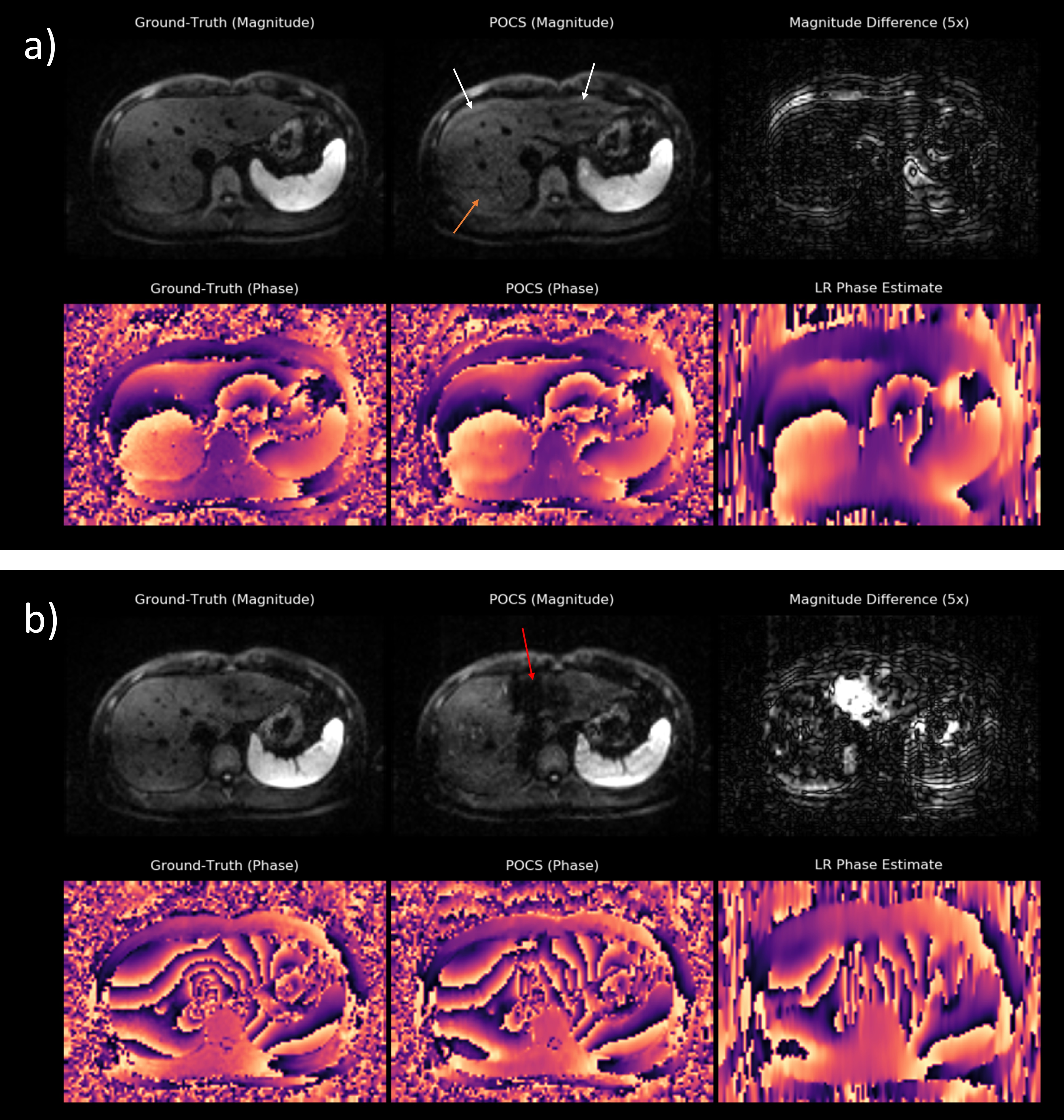
Figure 1: POCS
reconstructions of retrospectively PF-sampled liver images (b = 800 s/mm2) in two representative cases: a) one with
relatively smooth phase and b) one with high-frequency phase fluctuations. In
a), the phase can be approximately restored using the LR phase estimate shown
in the bottom right. However, ringing (white arrows) as well as a grid pattern (orange
arrow) are still introduced into the magnitude image. The artifacts in b) are
much more severe leading to
signal-dropout in the liver (red arrow) whose location coincides with wrong estimations of the phase.