lu wang1, Tian Li2, Jing Cai2, and Hing-Chiu Chang1
1Department of Diagnostic Radiology, The University of Hong Kong, HongKong, China, 2Department of Health Technology and Informatics, The Hong Kong Polytechnic University, HongKong, China
1Department of Diagnostic Radiology, The University of Hong Kong, HongKong, China, 2Department of Health Technology and Informatics, The Hong Kong Polytechnic University, HongKong, China
4D-DW-Propeller-EPI showed better geometric accuracy and image quality than 4D-DW-EPI for resolving respiratory motion, and its improved geometric accuracy may benefit the application of MRI-guided abdominal radiotherapy.
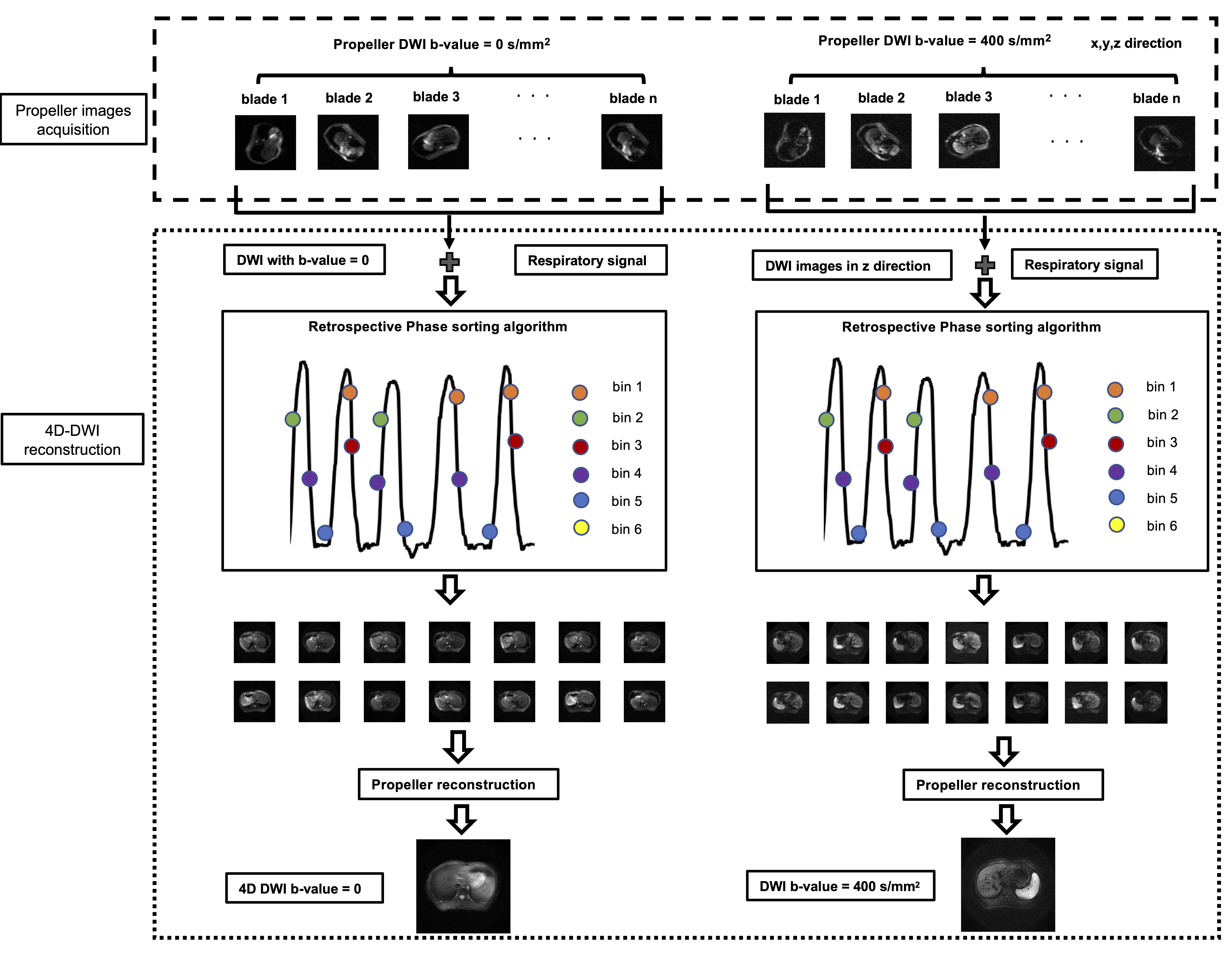
Fig.1 The framework of 4D-DW-Propeller-EPI technique with golden angle. It mainly includes two parts: blade data acquisition and 4D-DWI data reconstruction. Abdomen was scanned with 180 blades for each type of two diffusion contrast (b = 0 s/mm2 and b = 400 s/mm2). The recorded respiratory signal was used for sorting the blade data and placing them into corresponding phase bins. The blades falling into the same bin were combined using Propeller-EPI reconstruction.
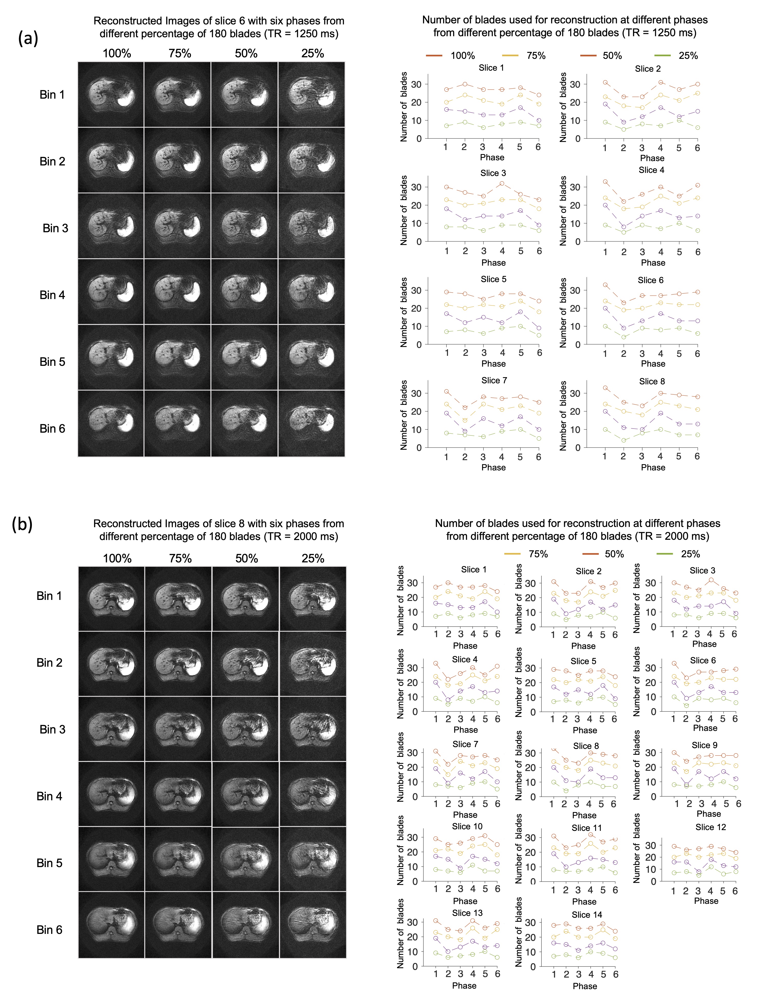
Fig.4 Left panel shows the In-vivo 4D-DW-Propeller-EPI images reconstructed from data acquired with (a) TR=1250ms, and (b) TR=2000ms. Right panel shows the number of blades used for reconstructing six respiratory phases for the usage of 100%, 75%, 50%, and 25% of all acquired blade data (180 blades). Shortening sampling duration can significantly reduce the number of blades available in each phase bin for subsequent Propeller-EPI reconstruction, resulting in degraded image quality. The reduction of blade number in different phases is not the same, leading to varying image quality.