Hiroshi Hamano1, Masami Yoneyama1, Akihiro Nishie2, Keisuke Ishimatsu2, Chiaki Tokunaga3, Hiroaki Watanuki3, Tatsuhiro Wada3, Isao Shiina4, Michinobu Nagao5, Yasuhiro Goto4, Kazuo Kodaira4, Yutaka Hamatani4, Takumi Ogawa4, Takashi Namiki1, and Kenji Iinuma1
1Philips Japan, Tokyo, Japan, 2Department of Clinical Radiology, Graduate School of Medical Sciences, Kyushu University, Fukuoka, Japan, 3Division of Radiology, Department of Medical Technology, Kyushu University Hospital, Fukuoka, Japan, 4Department of Radiological Services, Tokyo Women’s Medical University, Tokyo, Japan, 5Department of Diagnostic Imaging and Nuclear Medicine, Tokyo Women’s Medical University, Tokyo, Japan
1Philips Japan, Tokyo, Japan, 2Department of Clinical Radiology, Graduate School of Medical Sciences, Kyushu University, Fukuoka, Japan, 3Division of Radiology, Department of Medical Technology, Kyushu University Hospital, Fukuoka, Japan, 4Department of Radiological Services, Tokyo Women’s Medical University, Tokyo, Japan, 5Department of Diagnostic Imaging and Nuclear Medicine, Tokyo Women’s Medical University, Tokyo, Japan
Motion-Sensitive (MoSe) CINE
imaging, based on T2FFE, could clearly visualize the motion-insensitive cardiac
timing. We
demonstrated that the respiratory and cardiac trigged DWI with optimal cardiac
trigger delay using MoSe CINE imaging leads to the robustness of image quality
in liver DWI.
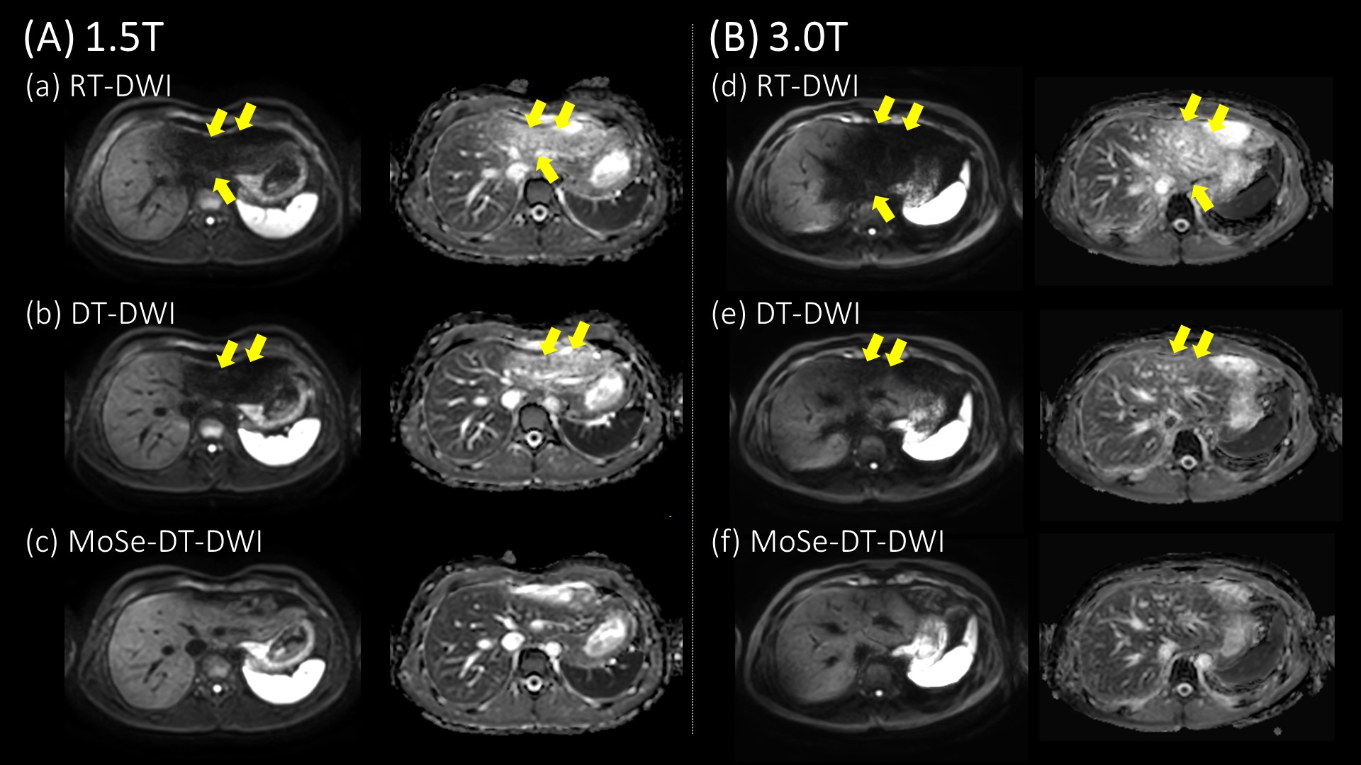
Figure 4. (A) Representative
RT-DWI and ADC map (a), DT-DWI and ADC map (b),
MoSe-DT-DWI
and ADC map (c) on a 1.5T scanner. In
the RT-DWI and DT-DWI, signal loss from cardiac related artifacts appeared and
the ADC left was artificially higher than ADC right (arrows). MoSe-DT-DWI
significantly improved the image qualities of the left liver lobe compared to
other techniques. (B) Representative RT-DWI and ADC map (d),
DT-DWI and ADC map (e), MoSe-DT-DWI and ADC map (f) on a 3T scanner, similar
findings to 1.5T were obtained.
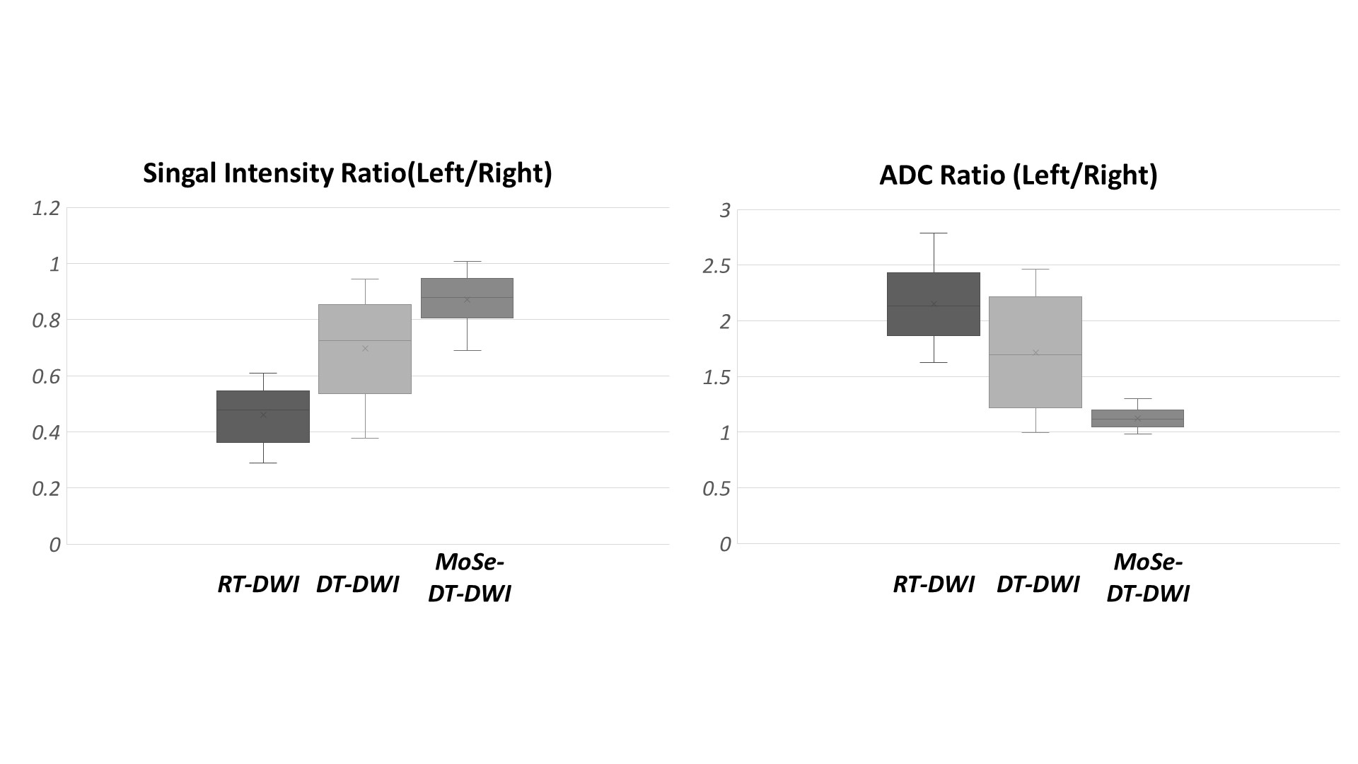
Figure 5. The SIRs and ADC ratios were shown. The
SIRs and ADC ratios at the MoSe-DT-DWI is the closest to one, suggests that MoSe-DT-DWI is the least impact of signal
loss from cardiac motion.