Hing-Chiu Chang1, Lu Wang1, Guangtao Chen1, Liyuan Liang1, Keith Wan-Hang Chiu1, Yi-Jui Liu2, Chun-Jung Juan3,4,5, and Hsiao-Wen Chung6,7
1Department of Diagnostic Radiology, The University of Hong Kong, Hong Kong, Hong Kong, 2Department of Automatic Control Engineering, Feng Chia University, Taichung, Taiwan, 3Department of Medical Imaging, China Medical University Hsinchu Hospital, Hsinchu, Taiwan, 4Department of Radiology, School of Medicine, College of Medicine, China Medical University, Taichung, Taiwan, 5Department of Medical Imaging, China Medical University Hospital, Taichung, Taiwan, 6Department of Electrical Engineering, National Taiwan University, Taipei, Taiwan, 7Graduate Institute of Biomedical Electronics and Bioinformatics, National Taiwan University, Taipei, Taiwan
1Department of Diagnostic Radiology, The University of Hong Kong, Hong Kong, Hong Kong, 2Department of Automatic Control Engineering, Feng Chia University, Taichung, Taiwan, 3Department of Medical Imaging, China Medical University Hsinchu Hospital, Hsinchu, Taiwan, 4Department of Radiology, School of Medicine, College of Medicine, China Medical University, Taichung, Taiwan, 5Department of Medical Imaging, China Medical University Hospital, Taichung, Taiwan, 6Department of Electrical Engineering, National Taiwan University, Taipei, Taiwan, 7Graduate Institute of Biomedical Electronics and Bioinformatics, National Taiwan University, Taipei, Taiwan
Free-breathing DW-Propeller-EPI can provide superior image quality for liver DWI, and
shows better repeatability in ADC measurement compared to three routine liver
DW-EPI methods.
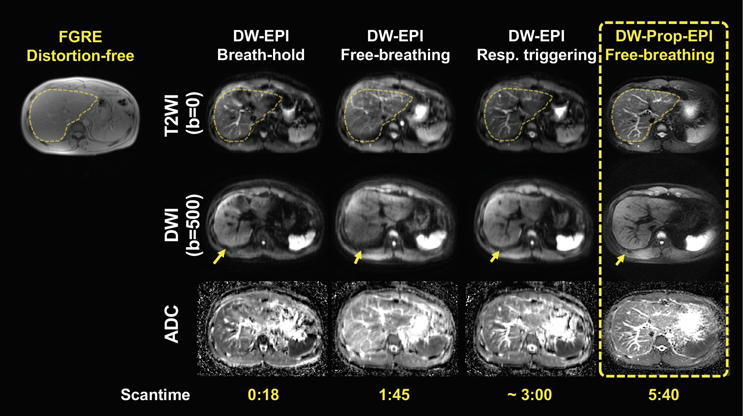
Fig.3 The representative liver diffusion
images (b=0 and b=500 s/mm2) and corresponding ADC maps generated
from three routine liver DW-EPI methods and free-breathing DW-Propeller-EPI.
Compared to three routine liver DWI methods, the DW-Propeller-EPI shows better
geometric accuracy (by comparing the liver contour obtained from FGRE image),
reduced geometric distortion (yellow arrows), and less image blurring. The
DW-Propeller-EPI is based on multi-shot acquisition, and therefore lengthens
the scantime (i.e., 5:40).
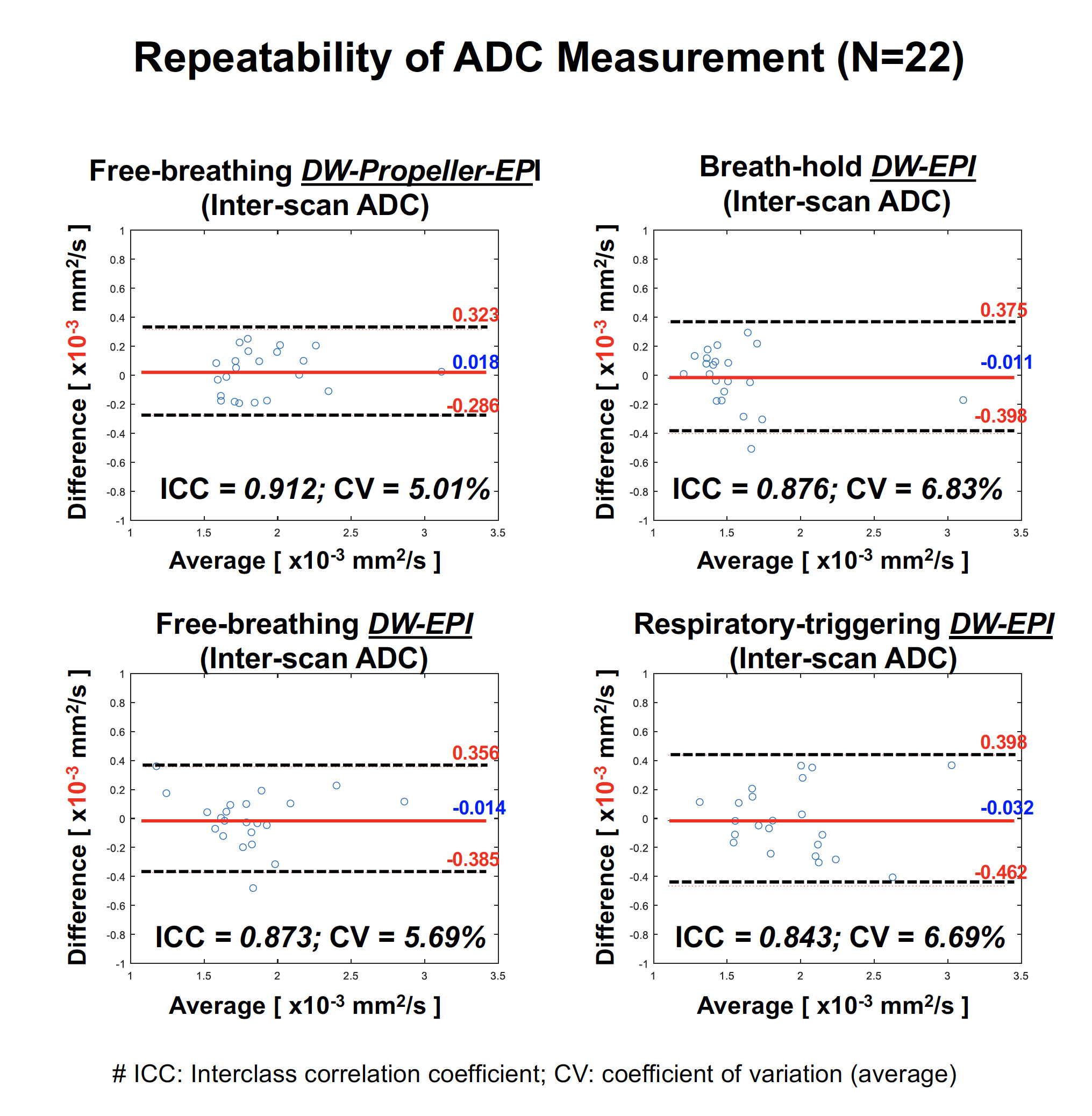
Fig.5 The evaluation of repeatability of ADC measurements of all sequences using the methods of
Bland and Altman. The free-breathing liver DW-Propeller-EPI shows best
repeatability in ADC measurement, with better ICC (0.912) and lower CV (5%)
than other three routine liver DW-EPI methods, suggesting that DW-Propeller-EPI
may be more suitable for cross-sectional
or longitudinal
liver DWI application.