Philip Meng-en Lee1, Jeremy W Gordon1, Zhen J Wang1, Zihan Zhu1, Hsin-Yu Chen1, Pamela N Munster2, Rahul Aggarwal2, Daniel B Vigneron1, and Michael A Ohliger1
1Department of Radiology & Biomedical Imaging, University of California, San Francisco, San Francisco, CA, United States, 2Department of Medicine, University of California, San Francisco, San Francisco, CA, United States
1Department of Radiology & Biomedical Imaging, University of California, San Francisco, San Francisco, CA, United States, 2Department of Medicine, University of California, San Francisco, San Francisco, CA, United States
Liver imaging with hyperpolarized 13C MRI is technically feasible with temporal resolution sufficient to detect distinct arterial and portal venous blood supplies. Understanding vascular dynamics in the liver will be crucial for timing and modelling of metabolic hyperpolarized signals.
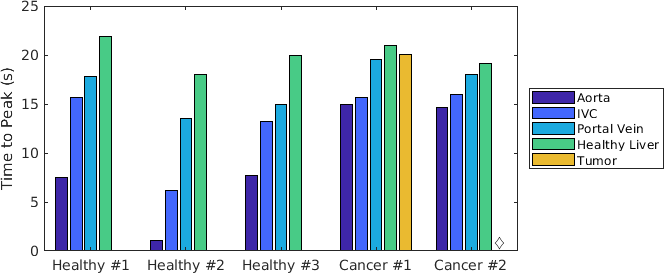
Figure 3. Peak arrival times of total 13C signal for each subject (t = 0 coincides with the start of the acquisition). The tumor for Cancer #2 fell within a saturation band and no signal was acquired (marked with a diamond).
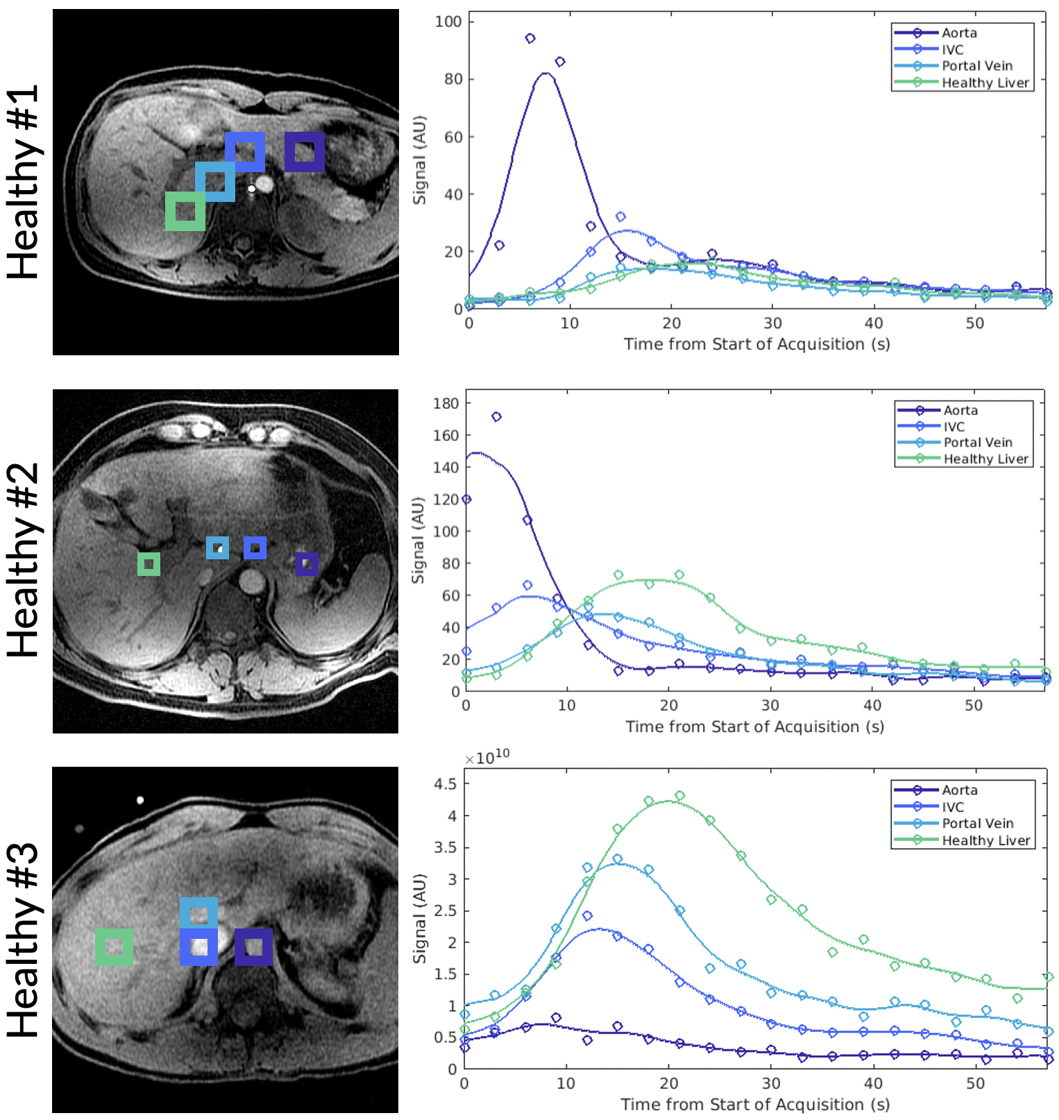
Figure 1. Total HP 13C signal plotted over time for selected tissue voxels in three healthy human subjects with ROIs labeled in the adjacent anatomical image. Spatial resolution varied between acquisitions.