Li An1, Shizhe Li1, Maria Ferraris Araneta1, Milalynn Victorino1, Christopher Johnson1, and Jun Shen1
1National Institute of Mental Health, National Institutes of Health, Bethesda, MD, United States
1National Institute of Mental Health, National Institutes of Health, Bethesda, MD, United States
A single-step
spectral editing 1H MRS technique with TE = 56 ms was used to measure fractional
enrichments of Glu and Gln in the human brain after oral administration of [U-13C]glucose.
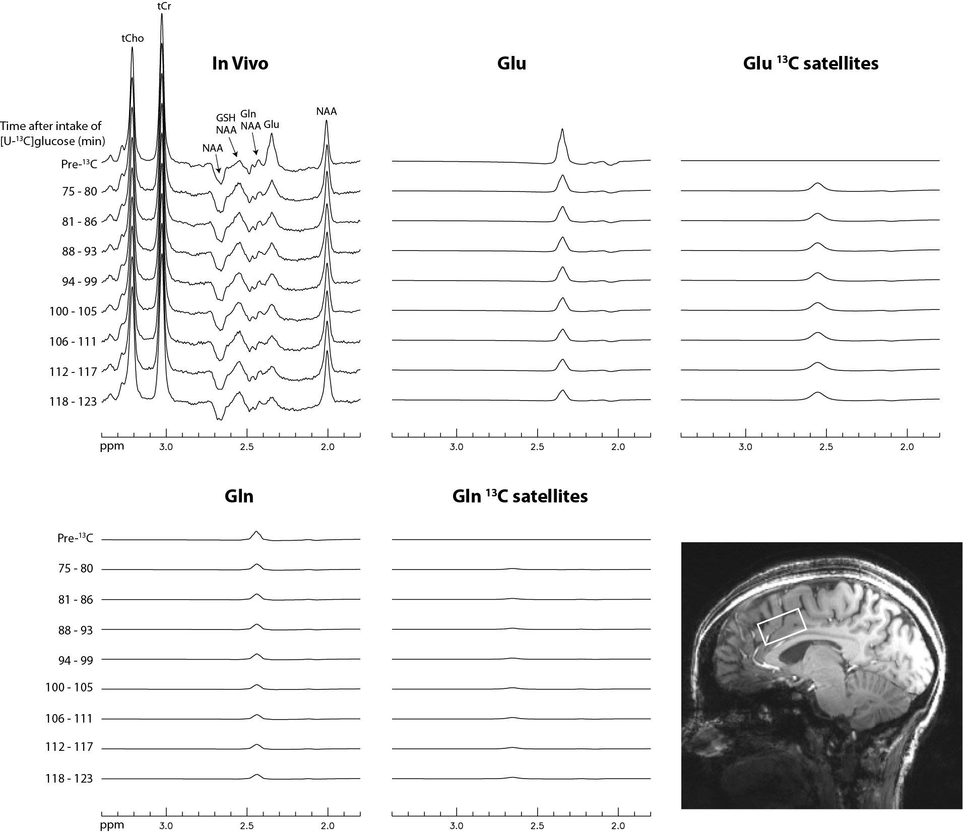
FIGURE 2 Time-course
spectra and corresponding fitted spectra of Glu, Gln, and their 13C
satellites acquired from the dACC of
subject 1. No linebroadening was applied to the spectra. Voxel size = 3.5 × 1.8 × 2 cm3; TR = 2.2 s; TE
= 56 ms; number of averages = 264 and total scan time = 10 min for the pre-13C
spectra; number of averages = 132 and total scan time = 5 min for each individual
post-13C
spectrum.
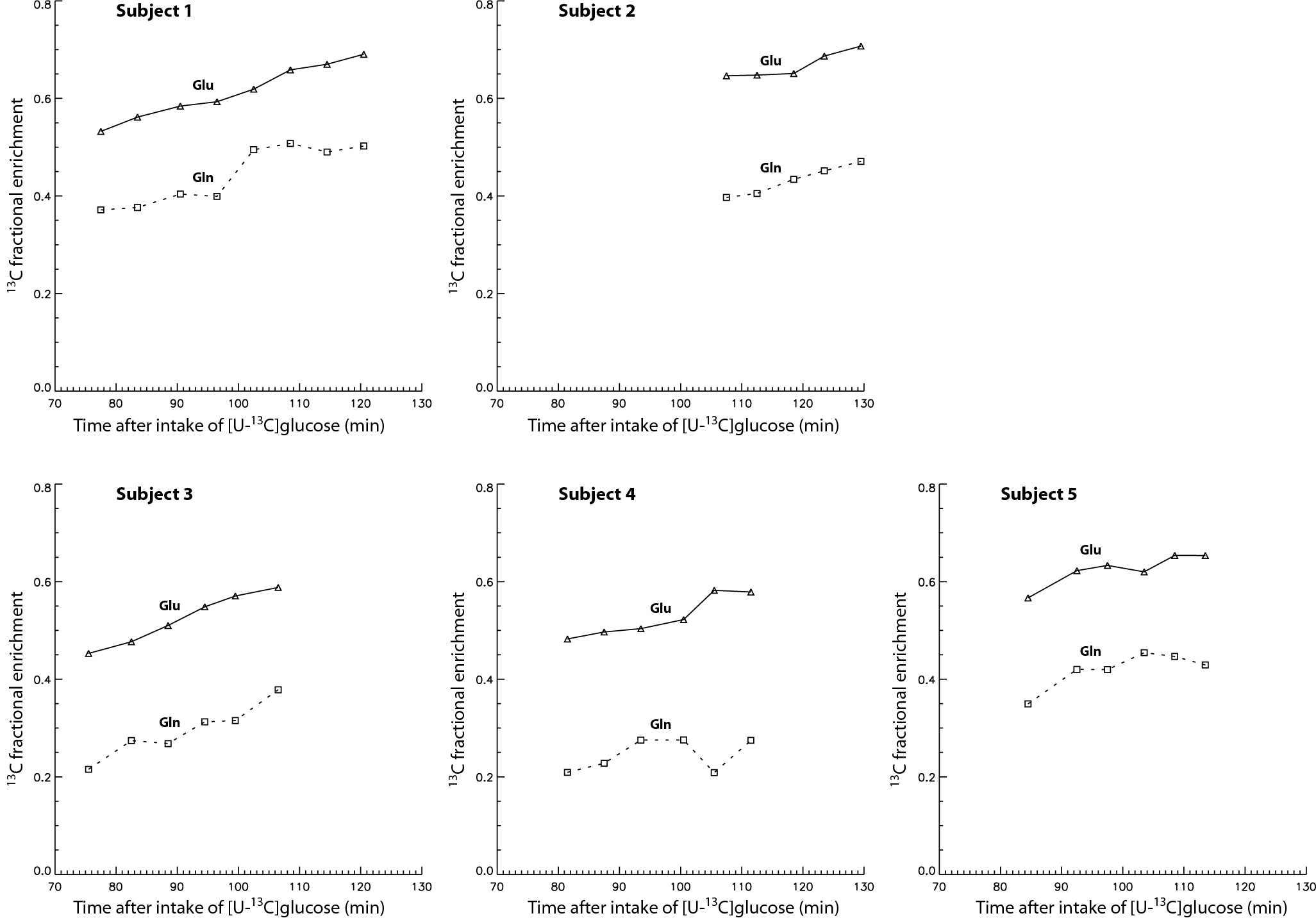
FIGURE 4 Plots of
fractional enrichments of Glu H4 and Gln H4 vs. time after oral
administration of [U-13C]glucose for all five healthy volunteers.