Steve C.N. Hui1,2, Tao Gong3, Helge J. Zöllner1,2, Yulu Song3, Yufan Chen3, Muhammad G. Saleh4, Mark Mikkelsen1,2, Georg Oeltzschner1,2, Sofie Tapper1,2, Weibo Chen5, Richard A.E. Edden1,2, and Guangbin Wang3
1Russell H. Morgan Department of Radiology and Radiological Science, The Johns Hopkins University School of Medicine, Baltimore, MD, United States, 2F.M. Kirby Research Center for Functional Brain Imaging, Kennedy Krieger Institute, Baltimore, MD, United States, 3Department of Imaging and Nuclear Medicine, Shandong Medical Imaging Research Institute, Cheeloo College of Medicine, Shandong University, Jinan, China, 4Department of Diagnostic Radiology and Nuclear Medicine, University of Maryland School of Medicine, Baltimore, MD, United States, 5Philips Healthcare, Shanghai, China
1Russell H. Morgan Department of Radiology and Radiological Science, The Johns Hopkins University School of Medicine, Baltimore, MD, United States, 2F.M. Kirby Research Center for Functional Brain Imaging, Kennedy Krieger Institute, Baltimore, MD, United States, 3Department of Imaging and Nuclear Medicine, Shandong Medical Imaging Research Institute, Cheeloo College of Medicine, Shandong University, Jinan, China, 4Department of Diagnostic Radiology and Nuclear Medicine, University of Maryland School of Medicine, Baltimore, MD, United States, 5Philips Healthcare, Shanghai, China
Metabolite-suppressed MM spectra were acquired in 100 subjects from 20 to 70 years of age. The MM spectra are strikingly similar between male and female subjects, and extremely stable as a function of age.
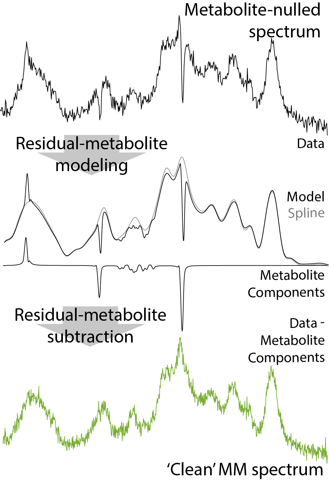
Figure 1. Osprey analysis workflow to remove residual metabolite signals and generate ‘clean’ MM spectrum.
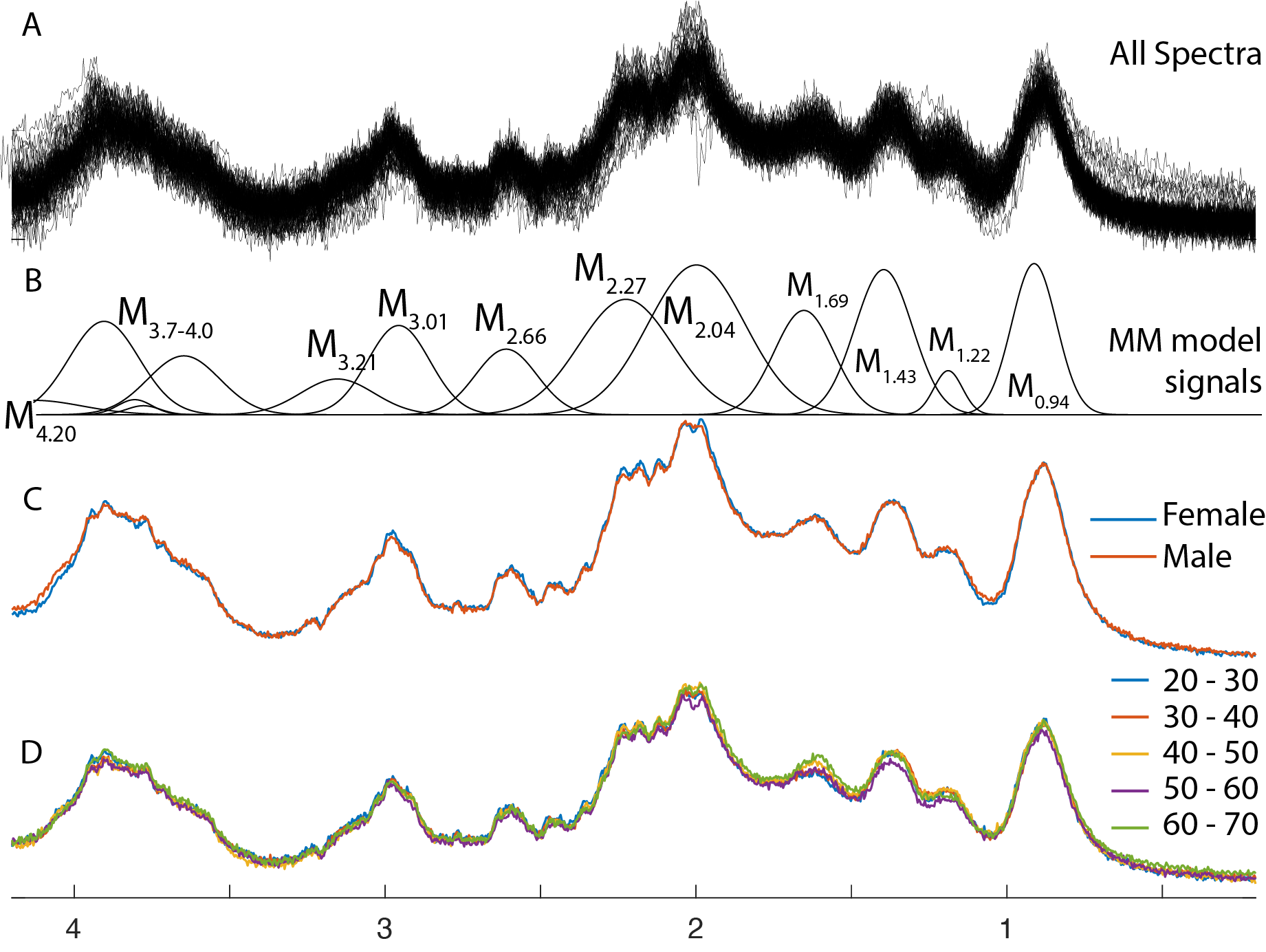
Figure 2. MM spectra from centrum semiovale white matter. Spectra from 96 subjects are overlaid in panel A, and the metabolite model signals are labeled in panel B. The mean spectra for male and female subjects are compared in panel C. The mean spectra for each decade of age are compared in panel D.