Wenqi Wang1, Xuan Jia2, Jiawei Liang2, Xiaohui Ma2, Weibo Chen3, Dan Wu1, Can Lai2, and Yi Zhang1
1Key Laboratory for Biomedical Engineering of Ministry of Education, Department of Biomedical Engineering, College of Biomedical Engineering & Instrument Science, Zhejiang University, Hangzhou, Zhejiang, China, 2Department of Radiology, Children’s Hospital, Zhejiang University School of Medicine, Hangzhou, Zhejiang, China, 3Philips Healthcare, Shanghai, China
1Key Laboratory for Biomedical Engineering of Ministry of Education, Department of Biomedical Engineering, College of Biomedical Engineering & Instrument Science, Zhejiang University, Hangzhou, Zhejiang, China, 2Department of Radiology, Children’s Hospital, Zhejiang University School of Medicine, Hangzhou, Zhejiang, China, 3Philips Healthcare, Shanghai, China
Amide proton transfer (APT) imaging was applied to evaluate the risk
of pediatric neuroblastoma in the abdomen. APT MRI was able to distinguish between low-
and high-risk abdominal NBs with an AUC of 0.917, which was substantially
higher than that of quantitative T1 and T2.
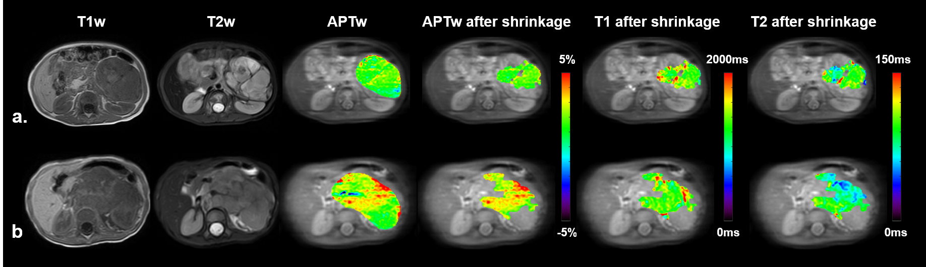
Figure
2. Conventional
anatomical images and APT-weighted images within initial ROI (third column) and
ROI after shrinkage with a cutoff value of 30% (fourth column), which were
fused with an image without saturation. Columns 5 and 6 show quantitative T1
and T2 maps calculated from MIX images within ROI after shrinkage, respectively.
Images were from a patient with low-risk neuroblastoma (a) and a patient with high-risk
neuroblastoma (b).

Table
2. The ROC analysis for differentiation
between low- and high-risk NBs