Xiaoxu Na1, Natalie E. Phelan1, Marinna R. Tadros1, Aline Andres2,3, Thomas M. Badger2,3, Charles M. Glasier1, Raghu H. Ramakrishnaiah1, Amy C. Rowell1, Li Wang4, Gang Li4, Zhengwang Wu4, David K. Williams5, and Xiawei Ou1,3,6
1Radiology, University of Arkansas for Medical Sciences, Little Rock, AR, United States, 2Pediatrics, University of Arkansas for Medical Sciences, Little Rock, AR, United States, 3Arkansas Children's Nutrition Center, Little Rock, AR, United States, 4Radiology, University of North Carolina at Chapel Hill, Chapel Hill, NC, United States, 5Biostatistics, University of Arkansas for Medical Sciences, Little Rock, AR, United States, 6Arkansas Children's Research Institute, Little Rock, AR, United States
1Radiology, University of Arkansas for Medical Sciences, Little Rock, AR, United States, 2Pediatrics, University of Arkansas for Medical Sciences, Little Rock, AR, United States, 3Arkansas Children's Nutrition Center, Little Rock, AR, United States, 4Radiology, University of North Carolina at Chapel Hill, Chapel Hill, NC, United States, 5Biostatistics, University of Arkansas for Medical Sciences, Little Rock, AR, United States, 6Arkansas Children's Research Institute, Little Rock, AR, United States
Significant differences in cortical thickness between infants born to
normal weight vs. obese mothers were found, and the cortical thickness was negatively correlated with maternal body fat mass percentage, suggesting impact of maternal obesity on offspring brain cortical development.
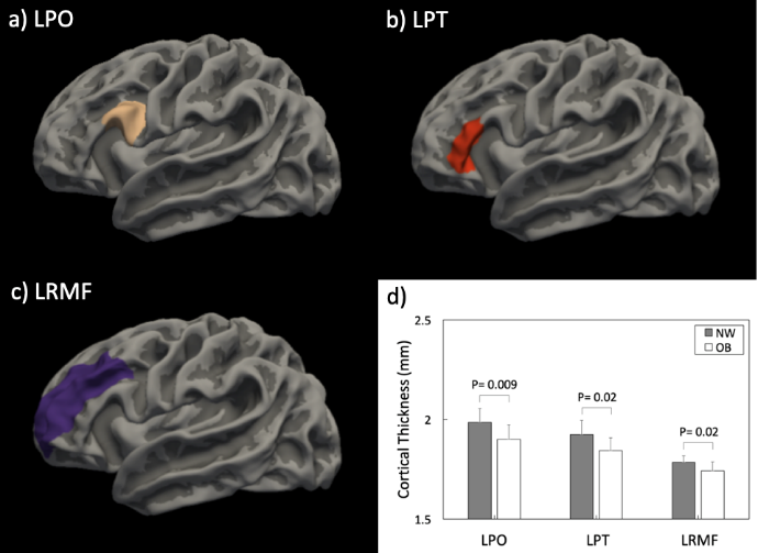
Figure 1: The anatomical locations for the 3 brain cortical regions
that showed significant differences (FDR corrected P < 0.05) in mean
cortical thickness between infants born to normal weight vs. obese pregnant
women: a) the left pars opercularis (LPO) gyrus; b) the left pars triangularis
(LPT) gyrus; and c) the left rostral middle frontal (LRMF) gyrus. The
comparison of mean cortical thickness for these 3 regions between these two
groups of infants is illustrated in d).
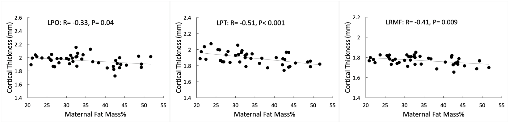
Figure 2:
Partial correlation analysis showed significant negative correlations (P <
0.05) between maternal body fat mass percentage measured at ~12 weeks of
pregnancy and infant brain mean cortical thickness measured at ~2 weeks of
postnatal age in the a) left pars opercularis (LPO) gyrus; b) left pars
triangularis (LPT) gyrus; and c) left rostral middle frontal (LRMF) gyrus.