Jacob Story1, Sevgi Gokce Kafali2,3, Shu-Fu Shih2,3, Kara L. Calkins4, Shahnaz Ghahremani3, and Holden H. Wu2,3
1David Geffen School of Medicine at UCLA, Los Angeles, CA, United States, 2Department of Bioengineering, University of California Los Angeles, Los Angeles, CA, United States, 3Department of Radiological Sciences, University of California Los Angeles, Los Angeles, CA, United States, 4Department of Pediatrics, University of California Los Angeles, Los Angeles, CA, United States
1David Geffen School of Medicine at UCLA, Los Angeles, CA, United States, 2Department of Bioengineering, University of California Los Angeles, Los Angeles, CA, United States, 3Department of Radiological Sciences, University of California Los Angeles, Los Angeles, CA, United States, 4Department of Pediatrics, University of California Los Angeles, Los Angeles, CA, United States
Free-breathing MR images were used to segment the pancreas on PDFF maps. In children with NAFLD, the pancreas has a high fat content that is predominantly in the superior regions. Pancreatic fat is related to BMI, liver fat, and acanthosis nigricans.
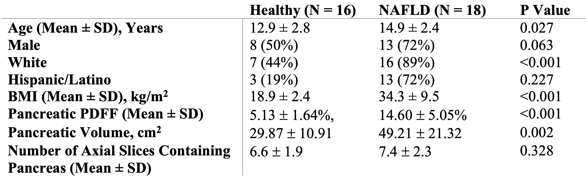
Table 1. Demographic Information of Study Participants. SD = Standard deviation. Body mass index (BMI), full segmentation pancreatic proton density fat fraction (PDFF) and pancreatic volume were significantly higher in children with nonalcohol fatty liver disease (NAFLD) than healthy children.
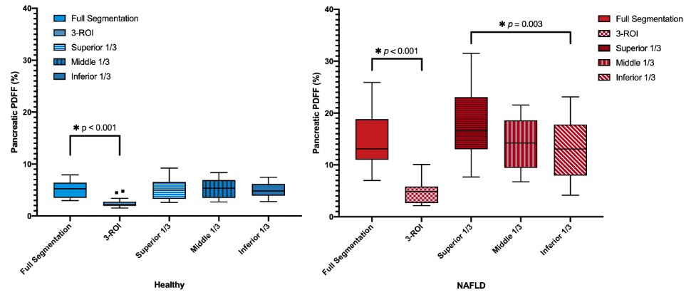
Figure 4. Box-Whisker Plots of Pancreatic Proton Density Fat Fraction (pPDFF) from Different Methods. Full segmentation (FS) pPDFF was higher in children with nonalcoholic fatty liver disease (NAFLD) compared to healthy children (p < 0.001). The 3-ROI method underestimated pPDFF compared to FS (p < 0.001 for healthy and NAFLD). The superior slices in children with NAFLD had higher pPDFF than inferior slices (p = 0.003).
