Shadi A Esfahani 1, Cody Callahan2, Nicholas M Rotile1, Peter D Caravan 1, Aaron K Grant 2, and Yi-Fen Yen1
1Athinoula A. Martinos Center for Biomedical Imaging, Department of Radiology, Massachusetts General Hospital, Boston, MA, United States, 2Department of Radiology, Beth Israel Deaconess Medical Center, Boston, MA, United States
1Athinoula A. Martinos Center for Biomedical Imaging, Department of Radiology, Massachusetts General Hospital, Boston, MA, United States, 2Department of Radiology, Beth Israel Deaconess Medical Center, Boston, MA, United States
Hyperpolarized [1-13C]Pyruvate Magnetic Resonance Spectroscopic Imaging is more sensitive for detection of early metabolic changes in gastric cancer after initiation of pan-RTK inhibition therapy compared to 18F-FDG PET/MRI, and can be used for prediction of response to novel therapies.
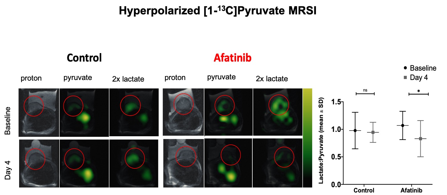
Fig 4. Hyperpolarized [1-13C]Pyruvate Magnetic Resonance Spectroscopic Imaging (MRSI) of the mice with NCI-N87 tumors (indicated by red circles) show significantly decreased lactate to pyruvate ratio (L/P) on day 4 of afatinib treatment compared to baseline, while the L/P ratio was not significantly changed in control group.
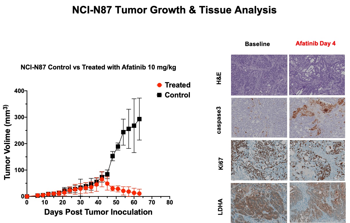
Fig 5. Treatment of the mice with sensitive NCI-N87 and resistant SNU16 tumors with afatinib showed no significant change in tumors size over 4 days of treatment in either group. Continued treatment in 3 weeks showed significant decrease in the size of NCI-N87 and significant growth of SNU16 tumors. Our in vivo HP-13C MRSI results were confirmed by immunohistochemistry evaluation of the tumor tissues; there is decreased anaerobic glycolysis marker, LDHA, decreased proliferation marker, Ki67, and increased apoptosis marker, Caspase3 in treated tumors compared to control.