Alexandra W. Acher1, Joseph Krenzer1, Krisztian Kovacs1, Soudabeh Kargar1, Ali Pirasteh1, Jitka Starekova1, TJ Colgan1, Victoria Rendell1, Daniel E. Abbott1, Erin Brooks1, Rashmi Agni1, Emily Winslow2, and Scott B. Reeder1
1University of Wisconsin School of Medicine and Public Health, Madison, WI, United States, 2Georgetown University, Washington DC, MD, United States
1University of Wisconsin School of Medicine and Public Health, Madison, WI, United States, 2Georgetown University, Washington DC, MD, United States
Radiologic histologic correlation of pancreas
cancer remains difficult. This feasibility study demonstrated successful
correlation of radiologic histologic pancreas cancer margins and features using
ex vivo MRI and a previously validated radiologic-histologic correlation
device.
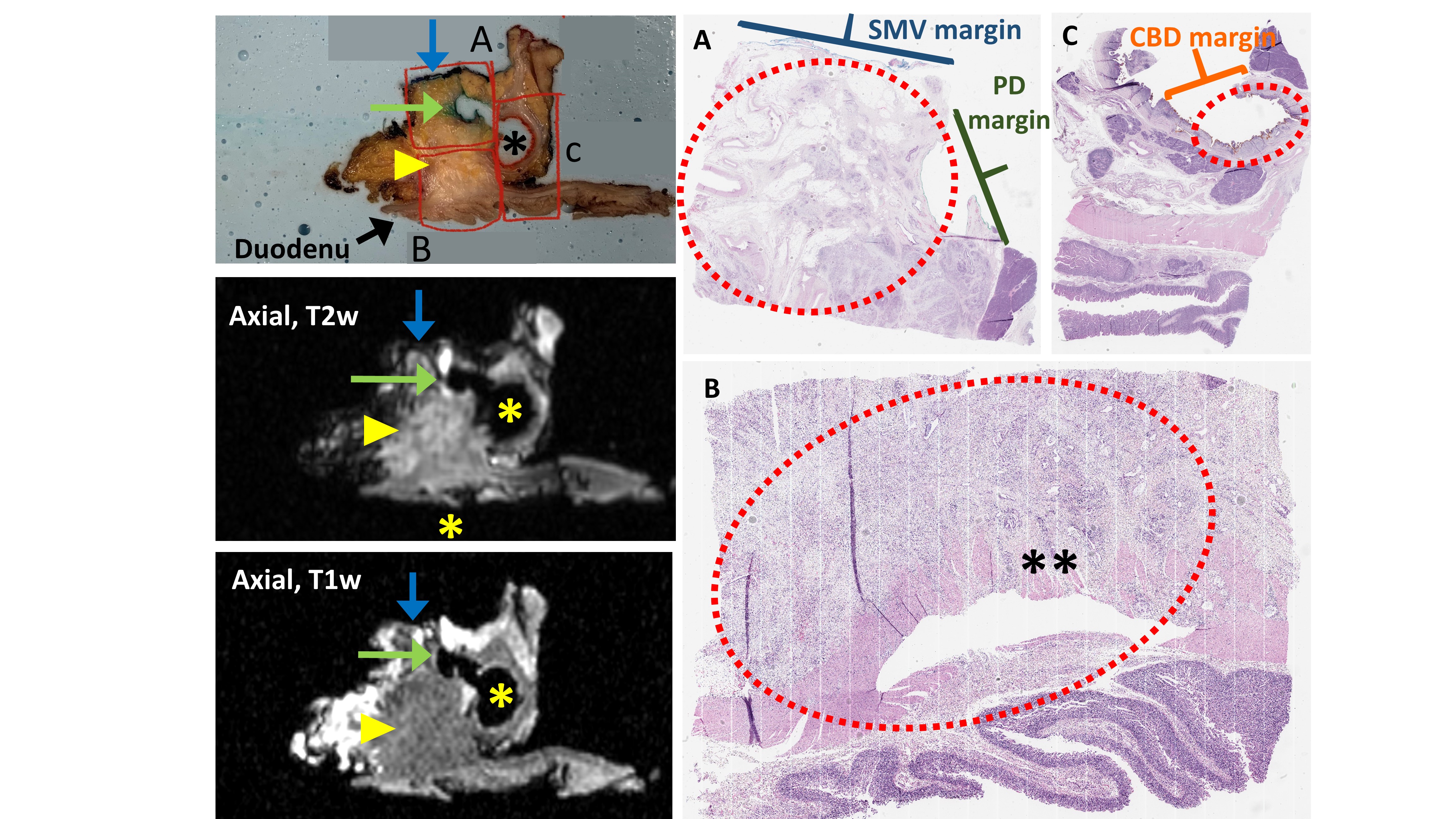
Counter clockwise from upper left: Axial cut of specimen with
alginate demonstrating mass (yellow triangle), SMV (blue arrow), CBD (*), PD
(green arrow) (red boxes define histology sections A-C); Corresponding
T2w axial slice with spiculated irregular mass with areas of hyper-intensity and
loss of fat plane between mass and duodenum (**); Corresponding T1w axial slice
demonstrating hypo-intense spiculated mass; Histology section A: tumor (red
circle), SMV and PD; B: tumor (red circle) with invasion into duodenal muscularis propria (**); C: CBD with tumor invasion (red circle).
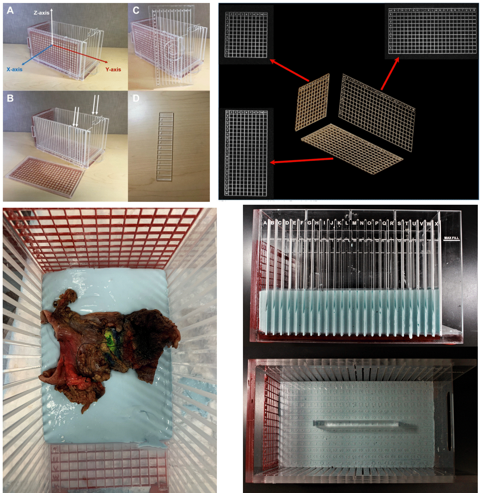
Counter clockwise from top left:
Radiologic histologic correlation device (RHCD) with
MRI visible grid depicting 3-dimensional axis and axial-orientation sectioning
grooves (white arrows); human pancreaticoduodenectomy specimen within alginate
(blue) in the RHCD; specimen submersion in alginate maintains orientation for
imaging and pathologic processing; 3D grid axis as depicted in MR images allows
for radiologic-histologic co-localization.