Leonie Petitclerc1,2, Lydiane Hirschler1, Jack A. Wells3, David L. Thomas4,5,6, and Matthias J.P. van Osch1,2
1C.J. Gorter Center for High Field MRI, Department of Radiology, Leiden University Medical Center, Leiden, Netherlands, 2Leiden Institute for Brain and Cognition (LIBC), Leiden, Netherlands, 3UCL Centre for Advanced Biomedical Imaging, Division of Medicine, University College London, London, United Kingdom, 4Neuroradiological Academic Unit, Department of Brain Repair and Rehabilitation, UCL Queen Square Institute of Neurology, University College London, London, United Kingdom, 5Dementia Research Centre, UCL Queen Square Institute of Neurology, University College London, London, United Kingdom, 6Wellcome Centre for Human Neuroimaging, UCL Queen Square Institute of Neurology, University College London, London, United Kingdom
1C.J. Gorter Center for High Field MRI, Department of Radiology, Leiden University Medical Center, Leiden, Netherlands, 2Leiden Institute for Brain and Cognition (LIBC), Leiden, Netherlands, 3UCL Centre for Advanced Biomedical Imaging, Division of Medicine, University College London, London, United Kingdom, 4Neuroradiological Academic Unit, Department of Brain Repair and Rehabilitation, UCL Queen Square Institute of Neurology, University College London, London, United Kingdom, 5Dementia Research Centre, UCL Queen Square Institute of Neurology, University College London, London, United Kingdom, 6Wellcome Centre for Human Neuroimaging, UCL Queen Square Institute of Neurology, University College London, London, United Kingdom
ASL signal
has been measured in the CSF of the human brain both in the ChP and around the
cortex. Preliminary modeling results point towards a transfer constant for
water to cross the BCSFB of ~100s.
Dynamic
representation of the blood, GM and CSF fractions of the ASL signal as
calculated with the previous parameter maps at arbitrary time points. The
labeling duration is 3s and the PLD is varied from 0 to 5s.
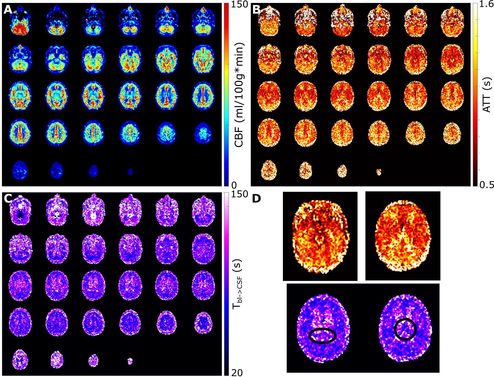
Parametric
maps obtained from voxel-wise fitting of the (smoothed) signal. The CBF (named
f in the dynamic model) and the ATT (or δ)
are shown in A and B. C shows the novel parameter Tbl->CSF, the
exchange time between the blood and CSF compartments. In D a close up of the
circle of Willis (top left) and a higher slice in the ATT map are given. The
bottom row of D present two slices in the Tbl->CSF map which show
low values in the choroid plexus (circled). Tbl->GM is not shown. The lowest slice of the imaging
volume crossed with the labeling plane which explains the discrepancy in
values.