Maria Marcella Lagana1, Noam Alperin2, Laura Pelizzari1, Ning Jin3, Domenico Zaca4, Marta Cazzoli1, Giuseppe Baselli5, and Francesca Baglio1
1CADiTeR, IRCCS Fondazione Don Carlo Gnocchi ONLUS, Milan, Italy, 2University of Miami, Miami, FL, United States, 3MR R&D Collaborations, Siemens Medical Solutions USA, Inc., Cleveland, OH, United States, 4Siemens Healthcare, Milan, Italy, 5Department of Electronics, Information, and Bioengineering, Politecnico di Milano, Milan, Italy
1CADiTeR, IRCCS Fondazione Don Carlo Gnocchi ONLUS, Milan, Italy, 2University of Miami, Miami, FL, United States, 3MR R&D Collaborations, Siemens Medical Solutions USA, Inc., Cleveland, OH, United States, 4Siemens Healthcare, Milan, Italy, 5Department of Electronics, Information, and Bioengineering, Politecnico di Milano, Milan, Italy
We measured the neck blood, and the cervical
cerebrospinal fluid flow rates using real-time phase contrast MRI. The power
spectra obtained from the flow rates had a peak at the breathing frequency, with
power increment from regular to deep breathing.
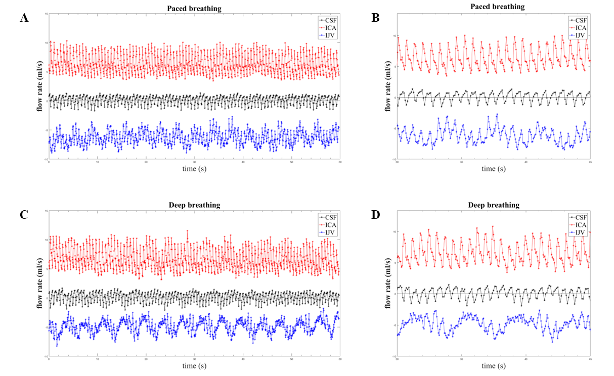
Figure 1. Temporal
curves of the ICAs, IJVs and CSF flow rates of a healthy volunteer in the paced
(A, B) and deep (C, D) conditions. The whole duration (60 s) is shown in A and
C, a sub-portion (seconds from 30 to 45) in B and D. Legend: ICA= internal
carotid artery; IJV= internal jugular vein; CSF=cerebrospinal fluid.
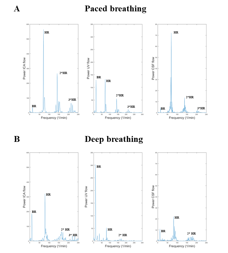
Figure 2. Amplitude spectra of internal
carotid arteries (ICAs), internal jugular veins (IJVs), cerebrospinal fluid (CSF)
flow rates (in (ml/s)2/Hz), separately for paced (A) and deep (B)
breathing. The lowest peak corresponds to the breathing frequency (BR) of the
subject, and increased its amplitude with deep breathing. Conversely, the heart
rate (HR) peak and its second (2*HR) and third (3*HR) harmonics decreased their
amplitudes with deep breathing.