Nivedita Agarwal1, Olivier Baledent2, Giuseppe Nicolò Frau3, and Sabino Walter Della Sala1
1Radiology, APSS Ospedale Santa Maria del Carmine, Rovereto, Italy, 2University of Amiens, Amiens, France, 3Otorhinolaryngology, APSS Ospedale Santa Maria del Carmine, Rovereto, Italy
1Radiology, APSS Ospedale Santa Maria del Carmine, Rovereto, Italy, 2University of Amiens, Amiens, France, 3Otorhinolaryngology, APSS Ospedale Santa Maria del Carmine, Rovereto, Italy
Meniere's disease is a disabling hearing disorder. Internal jugular venous stenosis may hamper overall neurofluid dynamics. However in this first ever analysis, phase contrast MRI suggests that our cohort had a normal overall dynamics likely due to efficient collateral venous flow.
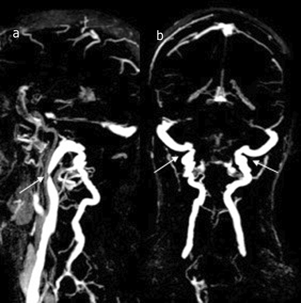
a) stenosis in the upper segment of the right IJV (white arrow). b) the presence of robust posterior and lateral condylar and deep cervical veins (double white arrows).
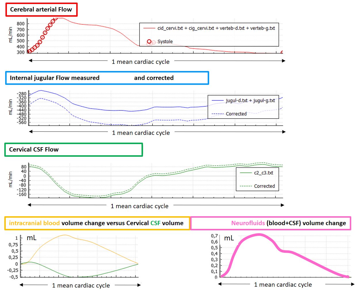
The curves represent overall neurofluid dynamics during cardiac cycle (CC). Cerebral arterial flow (red) (carotid and vertebral), IJV I(blue) was corrected (dotted line) to equal cerebral arterial flow. Intracranial blood volume change during CC (yellow), intracranial CSF volume change (green). Note how CSF responds to the intracranial blood pulsation. Nevertheless, CSF volume doesn’t fully compensate the blood volume and a net intracranial volume change is seen (pink).