Stephanie D Williams1, Nina E Fultz2, Nicole Tacugue3, Zenia Valdiviezo3, and Laura D Lewis2
1Psychological and Brain Sciences, Boston University, Boston, MA, United States, 2Biomedical Engineering, Boston University, Boston, MA, United States, 3Boston University, Boston, MA, United States
1Psychological and Brain Sciences, Boston University, Boston, MA, United States, 2Biomedical Engineering, Boston University, Boston, MA, United States, 3Boston University, Boston, MA, United States
Across three experiments, we show it is possible to induce CSF flow in the human brain during the awake state by manipulating hemodynamics with visually-evoked neural activity.
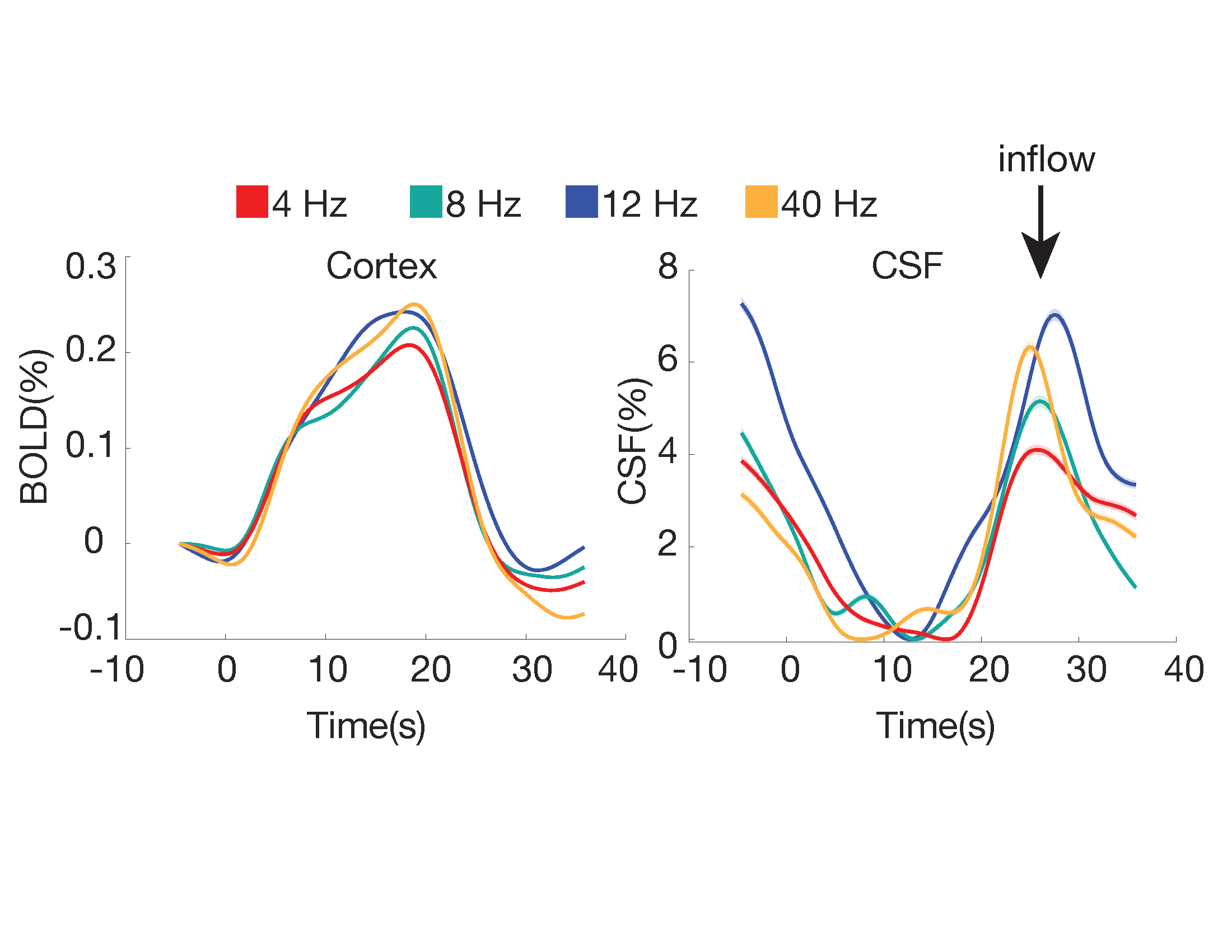
Figure 3: CSF inflow amplitude depends on visual stimulus frequency. The amplitude of the cortical BOLD (left panel) and CSF flow (right panel) responses to the visual stimulus each depend on checkerboard flicker frequency, with the largest responses to 12 Hz and 40 Hz. Shading indicates standard error; arrow indicates the timing of the CSF inflow event.
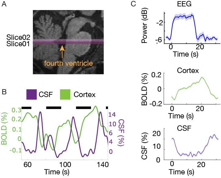
Figure 1: CSF flow is locked to the visually-evoked BOLD cortical signal (N=6). A) Example positioning of the functional image. B) Example smoothed cortical (green) and CSF (purple) traces from a single subject. Black bars indicate when the stimulus was on. The signals show consistent anticorrelated responses, with upwards CSF flow when the BOLD signal declines. C) The mean neural, hemodynamic and CSF signals across subjects shows that neural activity precedes hemodynamic and CSF responses.