Marco Muccio1, David Chu2, Lawrence Minkoff2, Neeraj Kukarni2, Brianna Damadian2, Raymond Damadian2, and Yulin Ge1
1Department of Radiology, New York University Grossman School of Medicine, New York City, NY, United States, 2FONAR Corporation, Melville, NY, United States
1Department of Radiology, New York University Grossman School of Medicine, New York City, NY, United States, 2FONAR Corporation, Melville, NY, United States
CSF exchange between the spinal canal and intracranial space is significantly increased when body position is shifted from upright to supine measured with a flow sensitive technique on a upright MRI.
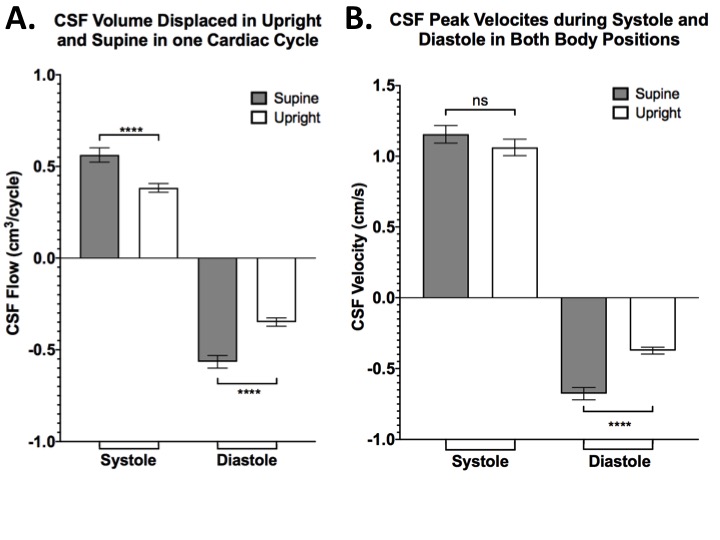
Figure 3. (A) CSF flow as stroke volume exchanged during one cardiac phase (systole or diastole) in upright and supine postures. The positive values indicate cranio-caudal direction while negative values indicate caudo-cranial direction. (B)CSF peak velocity measured during diastole and systole in supine and upright. Notice the difference in CSF peak velocity observed between supine and upright in diastole (****=p<0.0001) but are not significant in systole (ns=not significant).
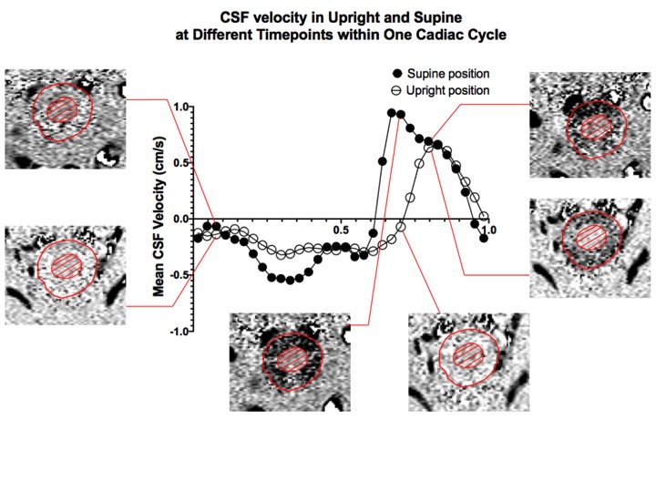
Figure 2. CSF flow rate at each time point over one cardiac cycle of a volunteer in the upright and supine posture at mid-C2. Note that the positive systolic peak (second half of the cardiac cycle) of the upright posture is only slightly reduced relative to the supine posture. Conversely, that of the negative upright diastolic peak (first half of the cardiac cycle) is reduced much more compared to the supine diastolic peak.