M. van den Kerkhof1,2, M.M. van der Thiel1,2, I.H.G.B. Ramakers2,3, R.J. van Oostenbrugge2,4,5, A.A. Postma1, A.A. Kroon5,6, J.F.A. Jansen1,2,7, and W.H. Backes1,2,5
1Department of Radiology & Nuclear Medicine, Maastricht University Medical Center, Maastricht, Netherlands, 2School for Mental Health & Neuroscience, Maastricht University, Maastricht, Netherlands, 3Department of Psychiatry & Neuropsychology, Maastricht University, Maastricht, Netherlands, 4Department of Neurology, Maastricht University Medical Center, Maastricht, Netherlands, 5School for Cardiovascular Disease, Maastricht University, Maastricht, Netherlands, 6Department of Internal Medicine, Maastricht University Medical Center, Maastricht, Netherlands, 7Department of Electrical Engineering, Eindhoven University of Technology, Maastricht, Netherlands
1Department of Radiology & Nuclear Medicine, Maastricht University Medical Center, Maastricht, Netherlands, 2School for Mental Health & Neuroscience, Maastricht University, Maastricht, Netherlands, 3Department of Psychiatry & Neuropsychology, Maastricht University, Maastricht, Netherlands, 4Department of Neurology, Maastricht University Medical Center, Maastricht, Netherlands, 5School for Cardiovascular Disease, Maastricht University, Maastricht, Netherlands, 6Department of Internal Medicine, Maastricht University Medical Center, Maastricht, Netherlands, 7Department of Electrical Engineering, Eindhoven University of Technology, Maastricht, Netherlands
Enabled by the recent developments at 7T MRI, this study identified a
relation between an increased blood flow velocity in the lenticulostriate
arteries and a higher number of enlarged perivascular spaces in the basal
ganglia and centrum semiovale.
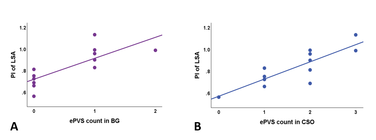
Figure 3. Scatterplots of PI measures in the LSA and ePVS score in the BG (A) and CSO (B). Least square linear regression
lines are added for visualization. ePVS = enlarged perivascular space, LSA = lenticulostriate arteries, BG = Basal Ganglia, CSO = Centrum Semiovale. ePVS
count is scored as follows: 0=<10 ePVS, 1=10-25 ePVS, 2=>25-40 ePVS,
3=>40 ePVS.
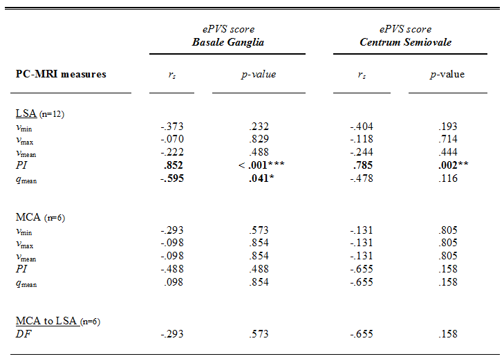
Table 2.
Spearman’s rho correlations between between ePVS count of the Basal
Ganglia and Centrum Semiovale and PC-MRI measures. Significant
associations are depicted in bold, ( * p < 0.05,
** p
< 0.01). PC-MRI = phase contrast magnetic resonance imaging, ePVS = enlarged
perivascular space, LSA = lenticulostriate arteries, MCA = middle cerebral artery, rs
= Spearman’s rho, v = blood flow velocity, q = volumetric flow
rate.