Muneeb A Faiq1, Vishnu Adi1, Anoop Sainulabdeen1, Sophia Khoja1, Carlos Parra1, Giles Hamilton-Fletcher1, Choong H Lee2, Jiangyang Zhang2, Gadi Wollstein1, Joel S Schuman1, and Kevin C Chan1,2
1Department of Ophthalmology, New York University Grossman School of Medicine, New York, NY, United States, 2Department of Radiology, New York University Grossman School of Medicine, New York, NY, United States
1Department of Ophthalmology, New York University Grossman School of Medicine, New York, NY, United States, 2Department of Radiology, New York University Grossman School of Medicine, New York, NY, United States
Using
contrast-enhanced MRI, the optic nerve appears to possess a waste clearance system via the cerebrospinal fluid in the paravascular space. This system appears molecular size-dependent and can
be modulated with aquaporin-4 water channel activity, concurring with the glymphatic system.
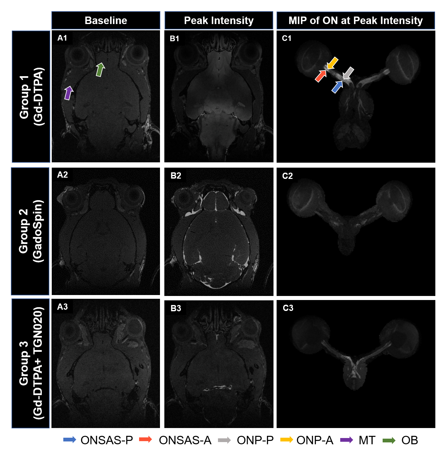
Figure
2. 3D dynamic
contrast-enhanced MRI at the level of the eye and the optic nerve
(ON) before
(A)
and
at peak intensity (B) after
Gd
infusion into
the subarachnoid space (SAS) of the lumbar spine. (C)
Maximum
intensity projection
(MIP) after
image segmentation of the eye and ON in
(B). Arrows indicate the regions of interest for quantitative analysis.
(ONSAS: optic nerve subarachnoid space;
ONP: optic nerve parenchyma; P: posterior; A: anterior; MT: muscle tissue; OB:
olfactory bulb)
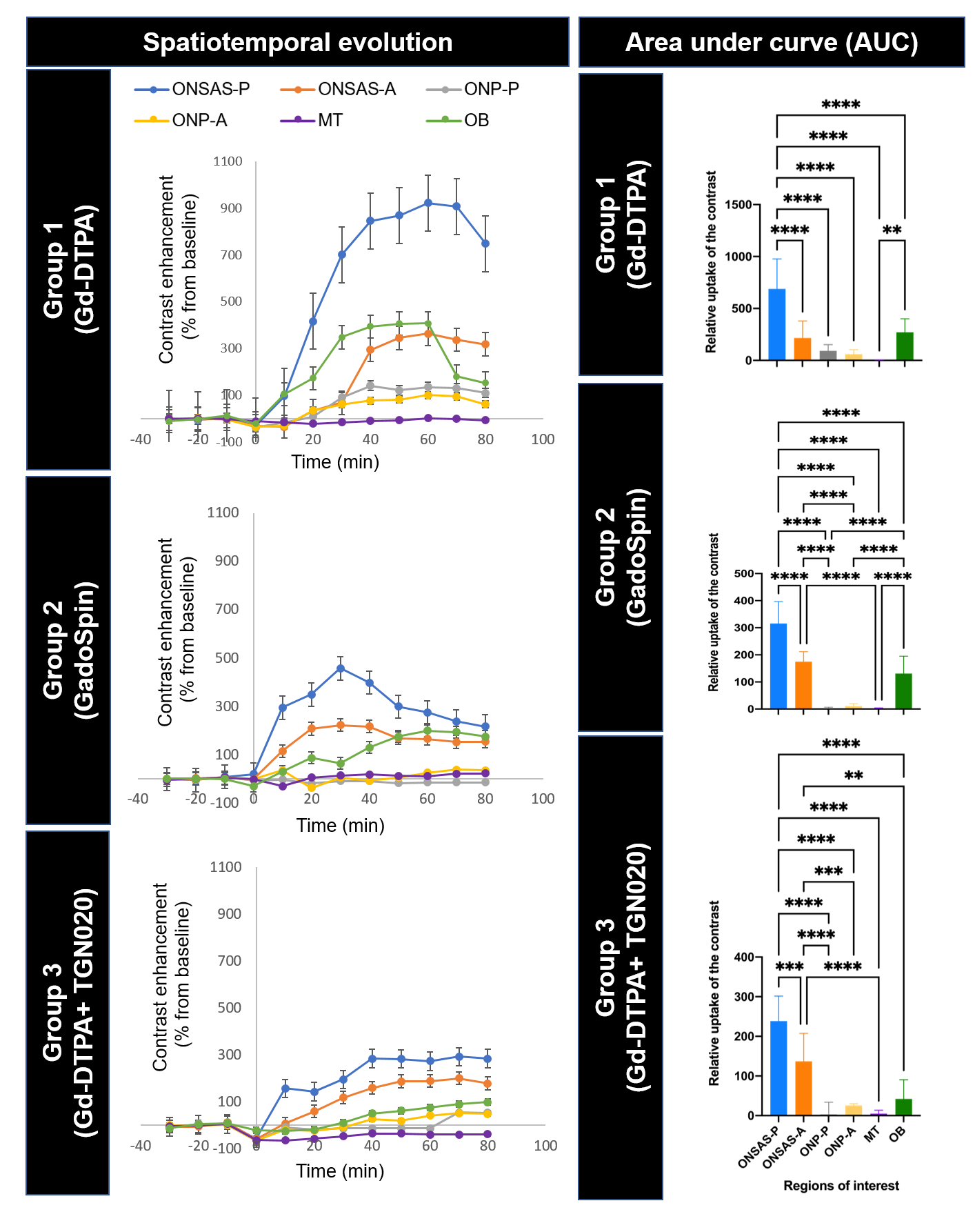
Figure
3: (Left column) Temporal
evolution of signal
enhancement
in the posterior optic nerve subarachnoid space (ONSAS-P), anterior ONSAS (ONSAS-A),
posterior
ON parenchyma (ONP-P),
anterior
ON parenchyma
(ONP-A), muscle
tissue
(MT), and olfactory bulb (OB) before
and after Gd contrast infusion into
SAS of the lumbar spine at timepoint zero. (Right
column) Relative
contrast uptake in
different regions of
interest using
area under curve (AUC). **p<0.01, ***p<0.001, ****p<0.0001.