Deneb Boito1,2, Magnus Herberthson3, Tom Dela Haije4, and Evren Özarslan1,2
1Department of Biomedical Engineering, Linköping University, Linköping, Sweden, 2Center for Medical Image Science and Visualization, Linköping University, Linköping, Sweden, 3Department of Mathematics, Linköping University, Linköping, Sweden, 4Department of Computer Science, University of Copenhagen, Copenhagen, Denmark
1Department of Biomedical Engineering, Linköping University, Linköping, Sweden, 2Center for Medical Image Science and Visualization, Linköping University, Linköping, Sweden, 3Department of Mathematics, Linköping University, Linköping, Sweden, 4Department of Computer Science, University of Copenhagen, Copenhagen, Denmark
Our results demonstrate the robustness of the QTI-derived parameters to data under-sampling when relevant positivity conditions are employed. The constrained estimation framework of QTI+ could make the technique feasible in clinical settings.
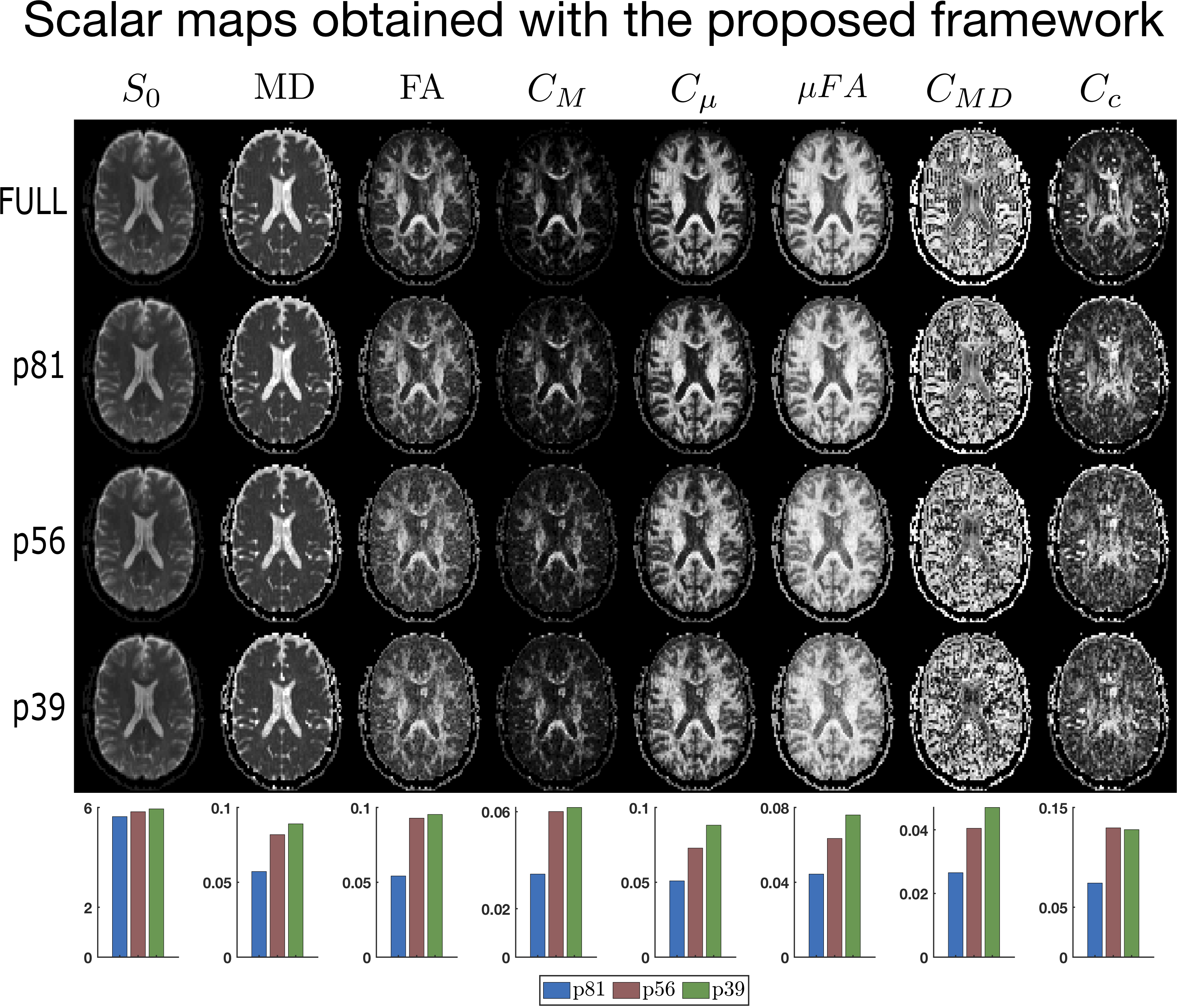
Figure 3. Scalar maps obtained by employing the QTI+ estimation on the full, p81, p56, and p39 protocols. Note how a large part of the information available in the full and p81 protocols is retained, when the p56 and p39 protocols were employed. The bars on the last row show the mean absolute deviation of the maps from their ground truth values (maps computed on the full dataset) for the three protocols. These plots highlight the resilience to downsampling achieved by imposing positivity constraints.
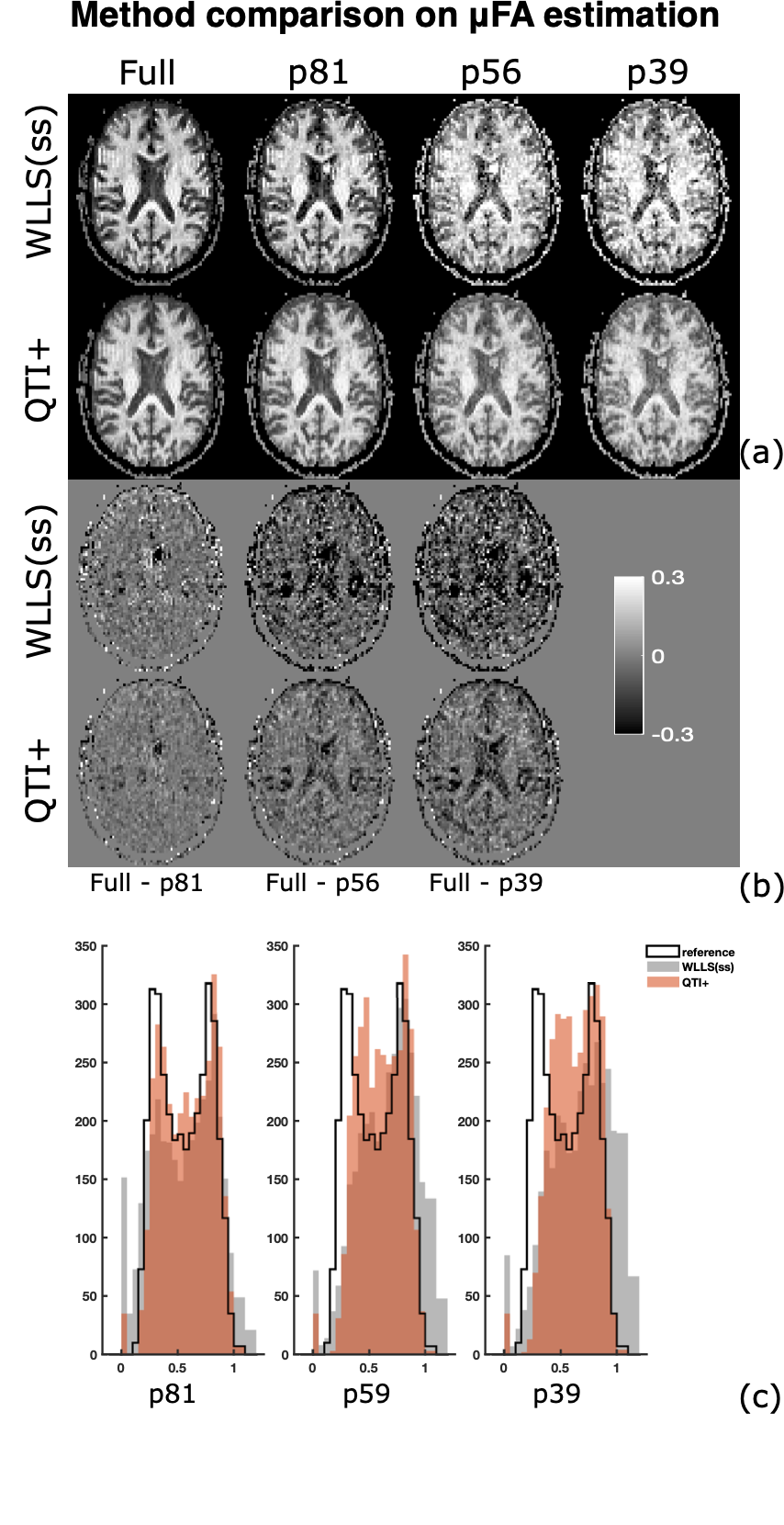
Figure 5. (a) $$$\mu FA$$$ maps obtained by fitting the QTI model with WLLS(ss) and emplying QTI+ on the four protocols. The fits performed with both methods on the full protocol are used as reference. (b) Difference between the reference $$$ \mu FA$$$ maps and those estimated with both methods on the three protocols. (c) Histograms showing the distribution of $$$ \mu FA$$$ values for the three protocols.