Eric Seth Michael1, Franciszek Hennel1, and Klaas Paul Pruessmann1
1Institute for Biomedical Engineering, ETH Zurich and University of Zurich, Zurich, Switzerland
1Institute for Biomedical Engineering, ETH Zurich and University of Zurich, Zurich, Switzerland
A diffusion imaging methodology combining enhanced
sensitivity OGSE waveforms with spiral readouts provided an improved depiction
of diffusion dispersion in the in-vivo human brain. The findings suggest the
relationship is better resolved at higher b-values.
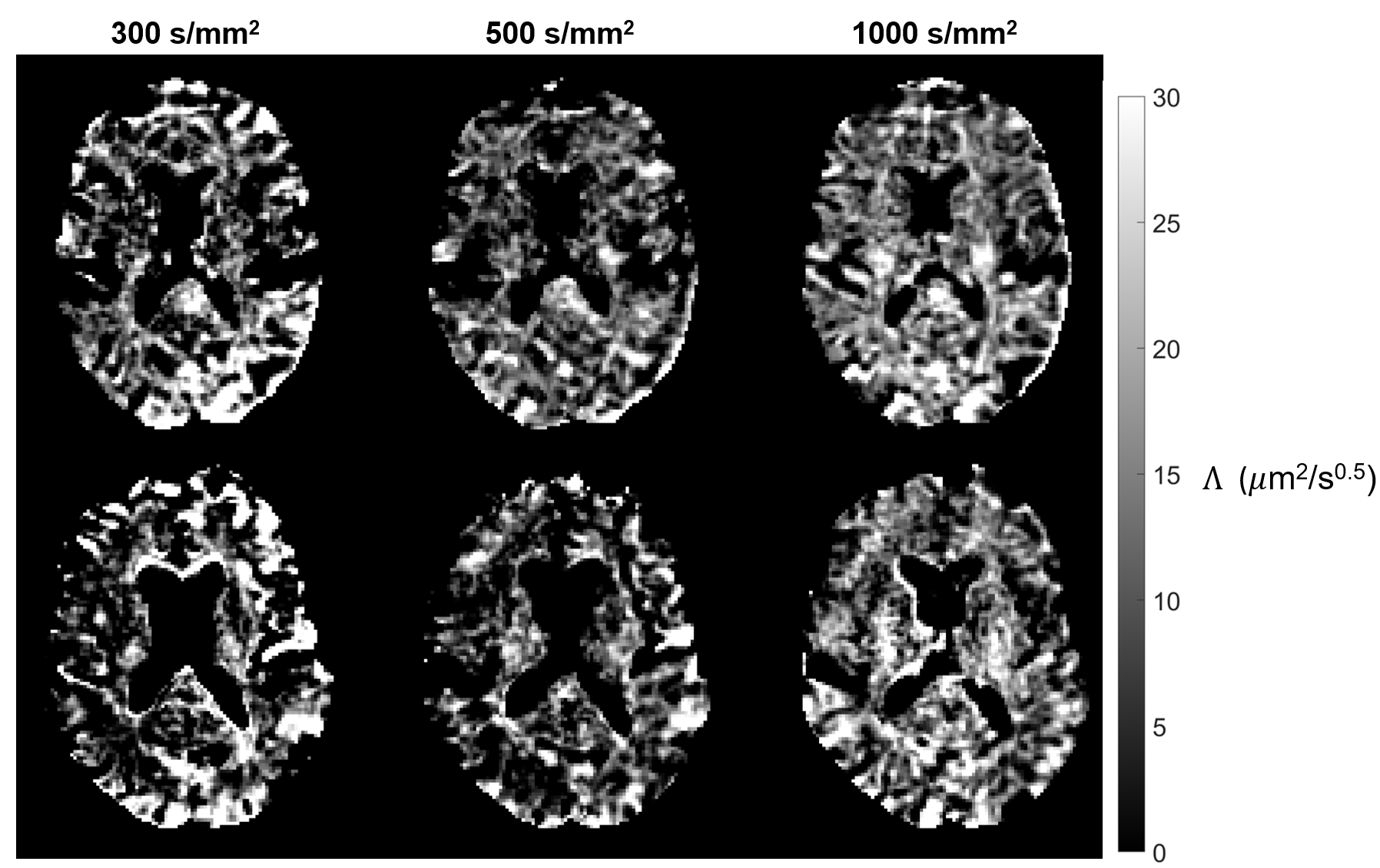
Figure 5. Diffusion dispersion rate (Λ) maps of a single slice from each subject (different rows) for all b-values (different columns), for OGSE shapes without flow compensation. All sampled frequencies for each respective b-value are accounted for in computation. Voxels with negative Λ are masked. The underlying contrast is similar for all b-values but stabilizes as the b-value increases (going from left to right), as fewer pixels exhibit spurious intensities.
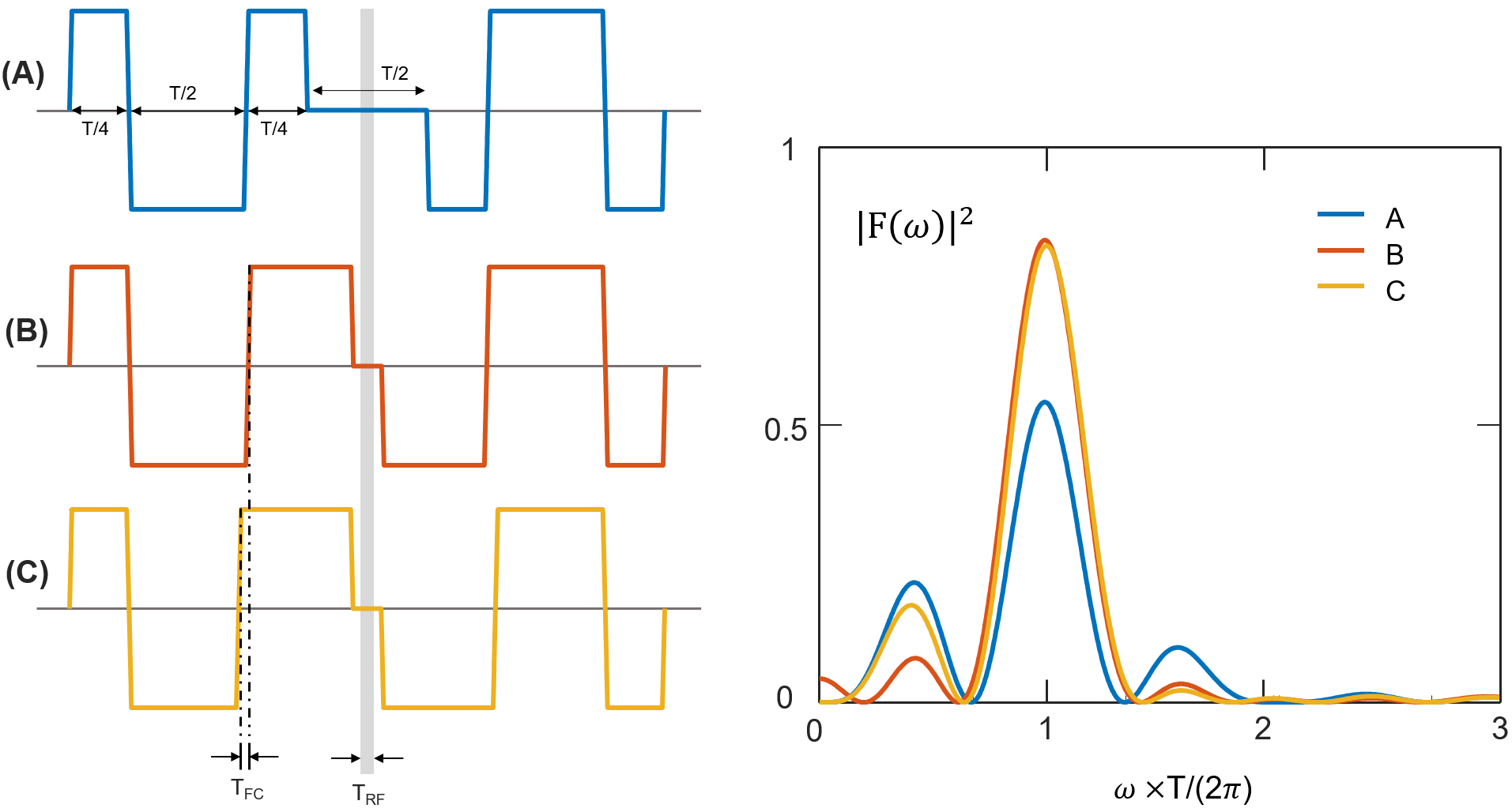
Figure 1. Comparison of OGSE gradient waveforms and power spectra. All plots are in arbitrary units, and the gradient plots show effective shapes including the inversion by the refocusing RF pulse. Power spectra are pictured for a standard OGSE shape with a half-period gap (A), a reduced gap OGSE shape2 (B), and a further modification to the latter producing FC (C). Note that the power spectra of (B) and (C) have higher peaks and smaller side lobes than that of (A). Also, the zero frequency peak of power spectrum (B) is eliminated in (C) by extending the innermost gradient lobes of (C) by TFC.
