Malwina Molendowska1, Fabrizio Fasano2,3, Umesh Rudrapatna1, Ralph Kimmlingen3, Derek K. Jones1,4, Slawomir Kusmia1, Chantal M. W. Tax1,5, and C. John Evans1
1Cardiff University Brain Research Imaging Centre (CUBRIC), Cardiff University, Cardiff, United Kingdom, 2Siemens Healthcare Ltd, Camberly, United Kingdom, 3Siemens Healthcare GmbH, Erlangen, Germany, 4Mary McKillop Institute for Health Research, Faculty of Health Sciences, Australian Catholic University, Melbourne, Australia, 5Image Sciences Institute, University Medical Center Utrecht, Utrecht, Netherlands
1Cardiff University Brain Research Imaging Centre (CUBRIC), Cardiff University, Cardiff, United Kingdom, 2Siemens Healthcare Ltd, Camberly, United Kingdom, 3Siemens Healthcare GmbH, Erlangen, Germany, 4Mary McKillop Institute for Health Research, Faculty of Health Sciences, Australian Catholic University, Melbourne, Australia, 5Image Sciences Institute, University Medical Center Utrecht, Utrecht, Netherlands
This study established the safety limits for the first ever operation of the 300 mT/m whole body system for body parts below the neck, and paves the way for unprecedented microstructural characterisation of heart and prostate tissue.
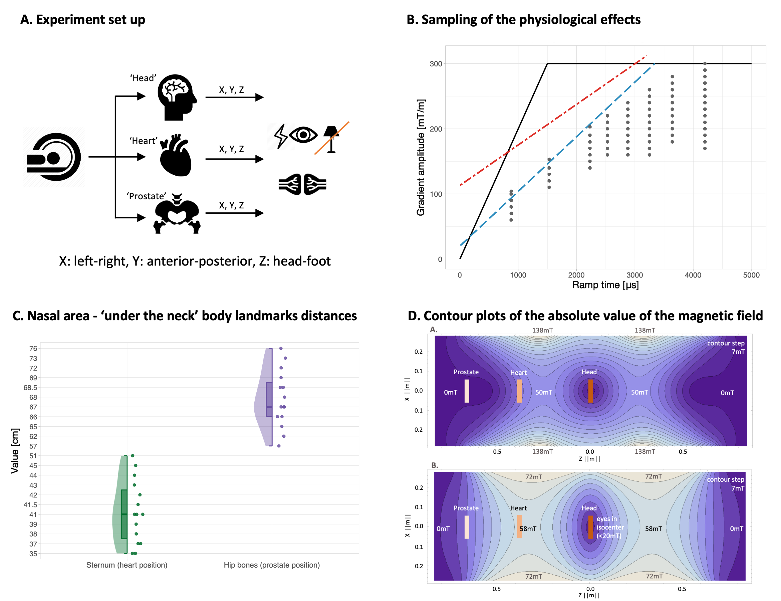
Fig. 1. A. Experimental set up. B. Acquisition protocol: dots show the sampling scheme within hardware (black), cardiac (red) or PNS (blue) limits for the Y-axis. C. Descriptive statistics of the measured relative distances of body landmarks. D. Contour plots of the absolute value of the magnetic field for a 300 mT/m gradient. The approximate positions of the eyes for prostate, heart or head landmarks are shown. Top row: |B| for a 300mT/m X-gradient (Y=0). Bottom row: |B| for a 300 mT/m Z-gradient (Y=0).
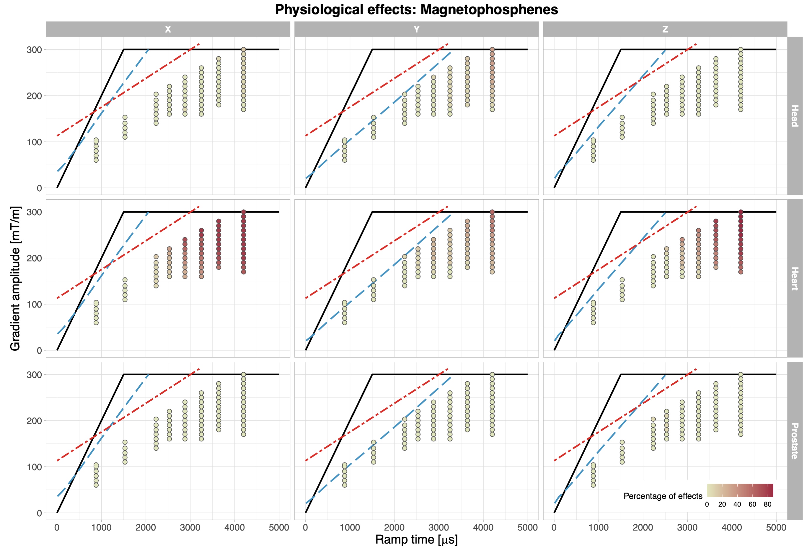
Fig. 3. 'Magnetophosphenes' occurrences for three imaging landmarks and three sampling axes. The dots are colour-coded according to the percentage of reported effects. The hardware limits are depicted as solid black curves, the cardiac stimulation limits as red dot-dashed lines, and the PNS limit as blue dashed lines, for X, Y, Z axes separately.