Mathias Davids1,2,3, Bastien Guerin1,2, and Lawrence L Wald1,2,4
1Martinos Center for Biomedical Imaging, Boston, MA, United States, 2Harvard Medical School, Boston, MA, United States, 3Computer Assisted Clinical Medicine, Mannheim, Germany, 4Harvard-MIT, Division of Health Sciences and Technology, Cambridge, MA, United States
1Martinos Center for Biomedical Imaging, Boston, MA, United States, 2Harvard Medical School, Boston, MA, United States, 3Computer Assisted Clinical Medicine, Mannheim, Germany, 4Harvard-MIT, Division of Health Sciences and Technology, Cambridge, MA, United States
Representing the PNS
information on a Huygens’ surface allowed assessment of the robustness of PNS
optimization in gradient design across body positions and models. Optimization
of a single position can retain PNS benefits for other body models and imaging
applications.
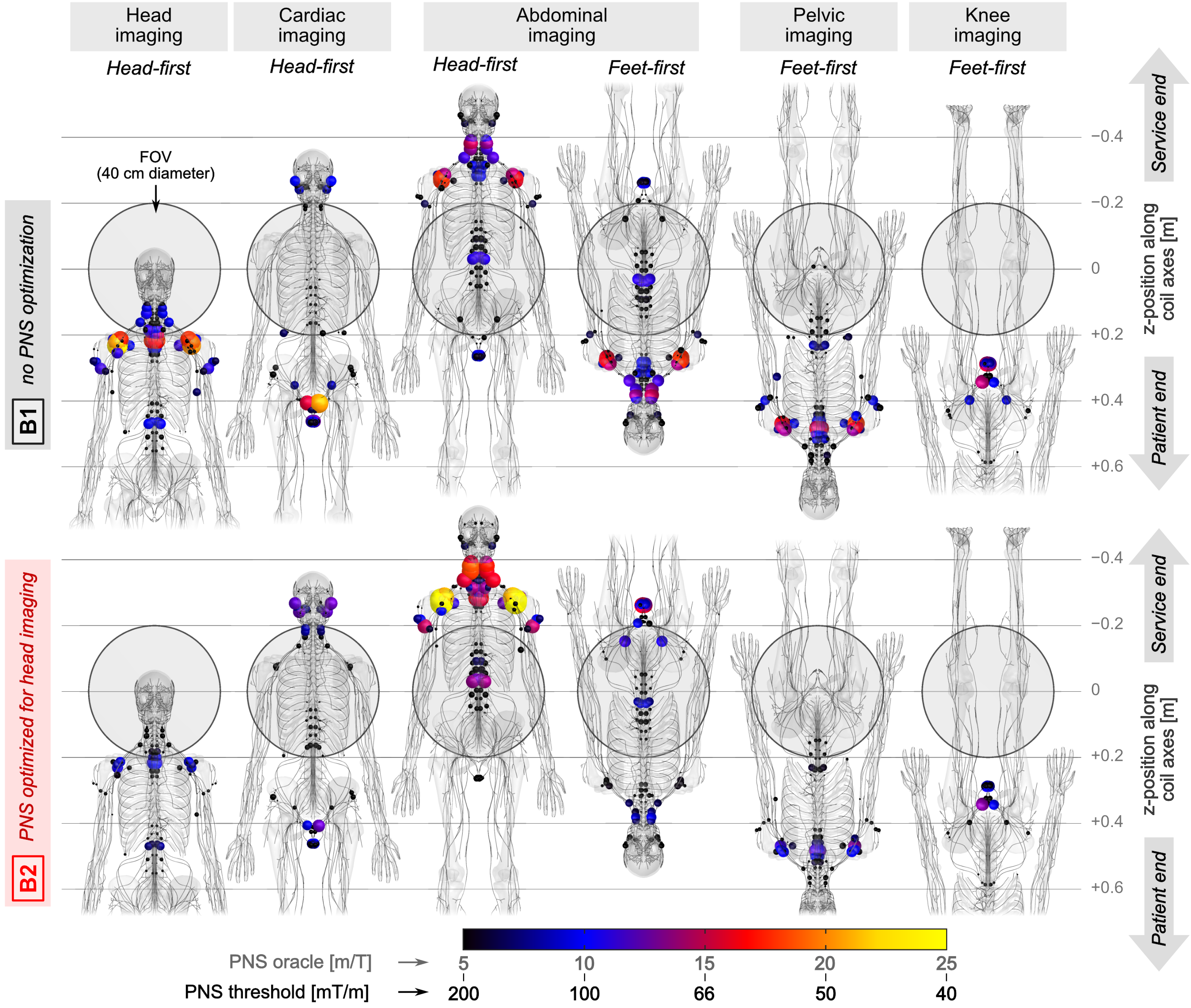
Figure 5: PNS hot-spots in the female model expected for
different body positions in the coils B1 (top) and B2 (bottom). Colored
spheres show hot-spots with largest PNS oracle (smallest PNS threshold). For abdominal
imaging, we show both head-first and feet-first supine poses; all
other scan positions use either head-first (head/cardiac imaging) or feet-first
pose (pelvic/knee imaging). In all cases, meant to correspond to conventional clinical
patient positions, the optimized coil retains some value in raising PNS
thresholds, except for the head-first supine abdominal imaging.
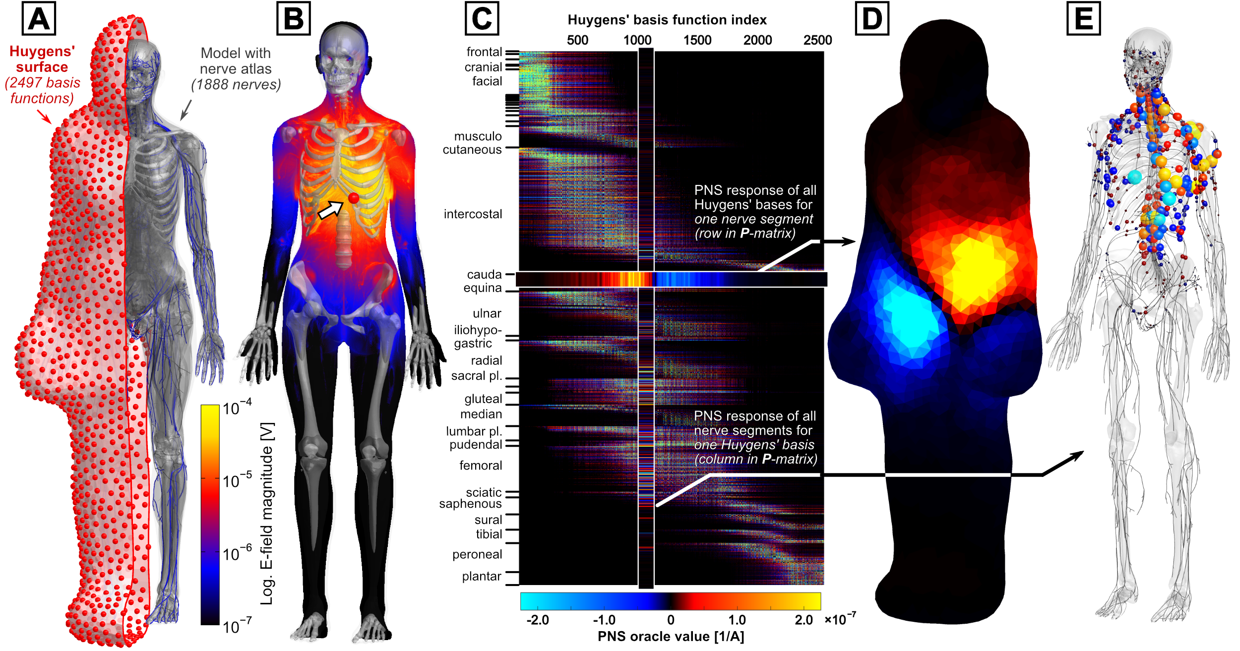
Figure 1: A: Definition
of the female model Huygens’ surface populated with 2497 magnetic basis
functions. B: E-field induced by
single basis near the heart. C:
After performing E-field
simulations and extracting PNS oracle values for all basis functions, we
assemble the Huygens’ P-matrix linking all 2497 bases (columns) to
PNS responses of all nerves (rows). D: PNS oracle for single 0.1 mm segment
of the cauda equina mapped onto the Huygens’ surface (highlighted row in P-matrix). E: PNS oracle of all nerve segments for a single Huygens’ basis
function (highlighted column in P-matrix).