Kellen Mulford1, David Darrow2, Sean Moen2, Samuel Ndoro2, Bharathi D. Jagadeesan3, Andrew W. Grande2, Donald R. Nixdorf4, and Pierre-Francois Van de Moortele1
1Center for Magnetic Resonance Imaging, University of Minnesota, Minneapolis, MN, United States, 2Department of Neurosurgery, University of Minnesota, Minneapolis, MN, United States, 3Department of Radiology, University of Minnesota, Minneapolis, MN, United States, 4Department of Diagnostic and Biological Science, University of Minnesota, Minneapolis, MN, United States
1Center for Magnetic Resonance Imaging, University of Minnesota, Minneapolis, MN, United States, 2Department of Neurosurgery, University of Minnesota, Minneapolis, MN, United States, 3Department of Radiology, University of Minnesota, Minneapolis, MN, United States, 4Department of Diagnostic and Biological Science, University of Minnesota, Minneapolis, MN, United States
This work establishes the feasibility of using 7.0-Tesla MRI to visualize acute and long-term treatment related effects from percutaneous radiofrequency rhizotomy procedures targeting the trigeminal ganglion.
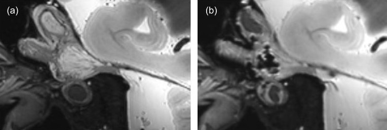
Figure 3: Sagittal views of (a) Pre-procedure 7.0-Tesla T2-SPACE imaging in the unpreserved human specimen shows high conspicuity in Meckel’s cave and the trigeminal ganglion. (b) The same location in the post-procedure imaging reveals widespread hypointensities.
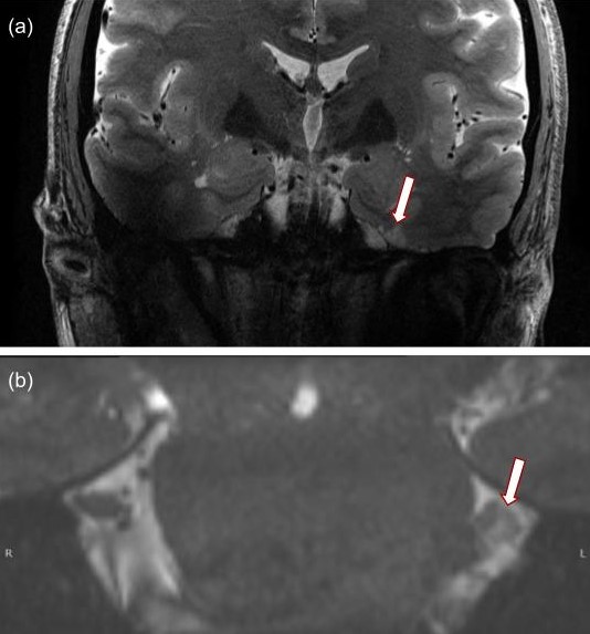
Figure 2: Coronal views of (a) Encephalomalacia on T2-TSE imaging and (b) nerve root atrophy on CISS imaging seen in a patient with four prior rhizotomy procedures at 7.0-Tesla.